. 2009 Mar 5;113(10):2324-35.
doi: 10.1182/blood-2008-03-146720. Epub 2008 Dec 22.

Differential requirement for the activation of the inflammasome for processing and release of IL-1beta in monocytes and macrophages

Affiliations

Differential requirement for the activation of the inflammasome for processing and release of IL-1beta in monocytes and macrophages

Mihai G Netea et al. Blood. .

Abstract

The processing of pro-interleukin-1beta depends on activation of caspase-1. Controversy has arisen whether Toll-like receptor (TLR) ligands alone can activate caspase-1 for release of interleukin-1beta (IL-1beta). Here we demonstrate that human blood monocytes release processed IL-1beta after a one-time stimulation with either TLR2 or TLR4 ligands, resulting from constitutively activated caspase-1 and release of endogenous adenosine triphosphate. The constitutive activation of caspase-1 depends on the inflammasome components, apoptosis-associated speck-like protein containing a caspase recruitment domain (ASC), and NALP3, but in monocytes caspase-1 activation is uncoupled from pathogen-associated molecular pattern recognition. In contrast, macrophages are unable to process and release IL-1beta solely by TLR ligands and require a second adenosine triphosphate stimulation. We conclude that IL-1beta production is differentially regulated in monocytes and macrophages, and this reflects their separate functions in host defense and inflammation.


Figures

Figure 1
Figure 1
Double-purified LPS is a specific TLR4 agonist. (A) Effect of LPS (1 μg/mL), PepG (10 μg/mL), and Pam3Cys (10 μg/mL) on surface expression of CD25 in CD14/TLR4-transfected CHO cells (relative flow cytometry units are shown). The results of 1 representative experiment of 3 are presented. (B) Mean ( ± SEM) TNF-α levels in supernatants from LPS (10 ng/mL)–stimulated PBMCs in the absence or absence of the TLR4 antagonist B quintana (100 ng/mL) indicated by aTLR4 (N = 5, P < .01, compared with LPS stimulation). (C) Effect of LPS (1 μg/mL), PepG (10 μg/mL), and Pam3Cys (10 μg/mL) on surface expression of CD25 in CD14/TLR2-transfected CHO cells (relative flow cytometry units are shown). The results of 1 representative experiment of 3 are presented. (D) Mean (± SEM) TNF-α levels in 24-hour supernatants of LPS-stimulated PBMCs from 5 volunteers bearing wild-type NOD2 allele (NOD2 WT) and 4 persons with a frame-shift mutation in NOD2 (NOD2fs).
Figure 2
Figure 2
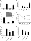
LPS induces bioactive IL-1β in primary monocytes. (A) Human PBMCs, purified CD14+ monocytes, or the CD14 lymphocytes were incubated with 10 ng/mL LPS, and the IL-1β concentrations were measured in the supernatant by specific ELISA after 24-hour incubation. (B) CD14+ monocytes were stimulated with various stimuli for the induction of IL-1β. (C) Human PBMCs were incubated with 10 ng/mL LPS and the levels of mature and precursor IL-1β or were measured in the supernatant by specific ELISA after 24-hour incubation. (Inset) Western blot of IL-1β and pro-IL-1β the supernatant derived from PBMCs stimulated with LPS (100 ng/mL) or MDP (10 μg/mL). (D) Dose-response of caspase-1 inhibitor ZVAD-fmk on the 24-hour levels of LPS-induced IL-1β from PBMCs. (E,F) The inhibitory effects of the caspase-1 inhibitor (20 μM) and of IL-1Ra (10 μg/mL) on the induction of IL-6 (E) and intracellular IL-1α measured in cell lysates (F) by 10 ng/mL LPS. Data from all 4 panels are presented as mean plus or minus SEM of cells harvested from 6 volunteers. *P < .05 compared with LPS stimulation alone.
Figure 3
Figure 3
Differential production of cytokines by monocytes, macrophages, and dendritic cells. Freshly isolated human PBMCs, macrophages differentiated after 5-day incubation with 10% human plasma, or dendritic cells differentiated after 5 days GM-CSF (50 ng/mL) + IL-4 (20 ng/mL), were stimulated with culture medium, LPS (100 ng/mL), Pam3Cys (10 μg/mL), or S epidermidis (106 organisms/mL). IL-1β (A,B), TNF (C), or IL-1α (D) was measured in the supernantant after 24-hour incubation. Data are presented as mean plus or minus SEM of cells harvested from 6 volunteers. *P < .01 compared with the stimulation in monocytes.
Figure 4
Figure 4
mRNA levels and processing of IL-1β in monocytes versus macrophages. (A) Steady-state levels of IL-1β and TNF-α in freshly isolated PBMCs or monocyte-derived macrophages after 4 hours of incubation with, LPS (100 ng/mL), Pam3Cys (10 μg/mL), or S epidermidis (106 organisms/mL). (B) Intracellular levels of precursor IL-1β in monocyte-derived macrophages stimulated for 24 hours with the various stimuli as in panel A. (C) Western blot of caspase-1 peptides in monocytes (Mo), macrophages (Mf), or THP-1 cells after 2 hours with and without LPS (100 ng/mL). (D) Western blot of the active p10 fragment of caspase-1 (sc515 antibody from Santa Cruz Biotechnology) in freshly isolated monocytes (column 1) and in monocytes that have adhered for 2 hours in polystyrene plates (column 2). (E) IL-1β and pro-interleukin-1β (proIL-1β) Western blots of lysates of monocyte-derived macrophages before and after stimulation for 24 hours with 100 ng/mL LPS.
Figure 5
Figure 5
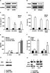
The role of ASC and NALP3 in the production and release of IL-1β. THP-1 cells were transfected with siRNA against ASC or NALP3 or control scramble (siC). RT-PCR of lysates after overnight incubation for ASC (A) or NALP3 (B). Cells were transfected with scrambled or target siRNA, then primed for 24 hours with PMA (100 ng/mL), and thereafter stimulated for 24 hours with LPS (1 μg/mL). After 24 hours, IL-1β (C) or IL-8 (D) was assessed in the supernatants (*P < .05 compared with the stimulation with LPS in cells treated with scramble siRNA). (E) Levels of supernatant IL-1β and TNF-α in primary PBMCs after transfection with scramble or siRNA for ASC and subsequent stimulation with LPS for 24 hours (n = 5, mean ± SEM, *P < .05 compared with LPS/scramble). (F) Levels of supernatant IL-1β in PBMCs stimulated for 3 hours with LPS (100 ng/mL), after which ATP (1 mM) or a combination of LPS and ATP was added for an additional 15 minutes. PBMCs were stimulated that were transfected with either scramble or siRNA against ASC (n = 5, means ± SEM, *P < .05 compared with LPS/ATP in the cells transfected with scramble siRNA). (G) Western blot of ASC in PBMCs transfected with either scramble or siRNA against ASC. (H) Western blot of the p10 and p45 caspase-1 in PBMCs transfected with either scramble (column 1) or siRNA (column 2) against ASC.
Figure 6
Figure 6
ATP induces IL-1β secretion in both monocytes and macrophages. (A) Human monocytes and macrophages were incubated with RPMI, LPS (1 μg/mL), or a combination of LPS and MDP (10 μg/mL) for 24 hours. Intracellular pro-IL-1β or secreted mature IL-1β were measured by specific ELISA kits (n = 6, mean ± SEM). (B) Monocytes or macrophages were incubated with RPMI or LPS for 4 hours, followed by ATP (1 mM) for 15 minutes (RPMI/ATP or LPS/ATP). Mean levels of mature IL-1β secreted in the supernatant were measured by ELISA (n = 6, mean ± SEM). (C) Western blots of caspase-1 in lysates after stimulation of monocytes with RPMI, LPS (1 μg/mL), ATP (1 mM), or a combination of LPS and ATP. (D) Monocytes and macrophages were incubated with RPMI or LPS (1 μg/mL) for 4 hours, and ATP was measured in the supernatant using a luciferase assay (n = 6, mean ± SEM, *P < .05 compared with monocytes). (E) Human monocytes and macrophages were incubated with RPMI or LPS (1 μg/mL) for 4 hours, and P2X7 receptors were blocked by adding oxidized ATP (oATP, 300 μM). Concentrations of mature IL-1β were measured in the supernatants by ELISA (n = 6, mean ± SEM, *P < .05 compared with RPMI). (F) Macrophages (Mf) differentiated in the presence of 10 ng/mL IFN-γ were stimulated for 24 hours with LPS (1 μg/mL), MDP (10 μg/mL), or heat-killed S epidermidis (106 organisms/mL). IL-1β in the supernatant was measured by ELISA (n = 6, mean ± SEM). (G) Macrophages differentiated for 5 days in RPMI with 10% plasma while in rotating (4 rpm) in 50-mL polypropylene tubes were stimulated for 24 hours with LPS, MDP, or heat-killed S epidermidis. IL-1β in the supernatant was measured by ELISA (n = 6, mean ± SEM). (H) Western blot of the active caspase-1 p10 fragment in macrophages differentiated in the absence (RPMI) or presence (IFN-γ) of IFN-γ. Data from 2 volunteers (1 and 2) are presented. (I) ATP release from macrophages differentiated in the absence or presence of IFN-γ (n = 5, mean ± SEM, *P < .05 compared with macrophages without IFN-γ).
Figure 7
Figure 7
Alveolar macrophages require the second stimulation by ATP to release mature IL-1β after LPS. (A) Human alveolar macrophages were stimulated for 24 hours with RPMI, LPS (100 ng/mL), Pam3Cys (10 μg/mL), or S epidermidis (106 organisms/mL). (B) Alveolar macrophages were stimulated with LPS (1 μg/mL) or a combination of LPS and ATP (1 mM). Intracellular pro-IL-1β after LPS stimulation after 4 hours, and the extracellular IL-1β release after stimulation with LPS or LPS/ATP, was assessed by ELISA. The stimulation experiments were performed in cells harvested from 5 volunteers (means ± SEM, *P < .05 compared with LPS stimulation alone). (C) Diagram representing the IL-1β activation pathways in monocytes and macrophages. Caspase-1 is constitutively activated in monocytes, and these cells release mature IL-1β after single stimulation with TLR ligands. IL-1β secretion is induced by endogenously released ATP. In contrast, macrophages need a double stimulation: one stimulus (TLR ligands) induces transcription, and a second stimulus (ATP) induces IL-1β secretion.

References

    1. Chae JJ, Wood G, Masters SL, et al. The B30.2 domain of pyrin, the familial Mediterranean fever protein, interacts directly with caspase-1 to modulate IL-1beta production. Proc Natl Acad Sci U S A. 2006;103:9982–9987. - PMC - PubMed
    1. Hoffman HM, Rosengren S, Boyle DL, et al. Prevention of cold-associated acute inflammation in familial cold autoinflammatory syndrome by interleukin-1 receptor antagonist. Lancet. 2004;364:1779–1785. - PMC - PubMed
    1. Hawkins PN, Lachmann HJ, Aganna E, McDermott MF. Spectrum of clinical features in Muckle-Wells syndrome and response to anakinra. Arthritis Rheum. 2004;50:607–612. - PubMed
    1. Aksentijevich I, Nowak M, Mallah M, et al. De novo CIAS1 mutations, cytokine activation, and evidence for genetic heterogeneity in patients with neonatal-onset multisystem inflammatory disease (NOMID): a new member of the expanding family of pyrin-associated autoinflammatory diseases. Arthritis Rheum. 2002;46:3340–3348. - PMC - PubMed
    1. van der Meer JW, Vossen JM, Radl J, et al. Hyperimmunoglobulinaemia D and periodic fever: a new syndrome. Lancet. 1984;1:1087–1090. - PubMed

Publication types

MeSH terms