. 2009 Jan;119(1):169-81.
doi: 10.1172/JCI35440. Epub 2008 Dec 22.

Abnormal expression of TIP30 and arrested nucleocytoplasmic transport within oligodendrocyte precursor cells in multiple sclerosis

Affiliations

Abnormal expression of TIP30 and arrested nucleocytoplasmic transport within oligodendrocyte precursor cells in multiple sclerosis

Jin Nakahara et al. J Clin Invest. 2009 Jan.

Abstract

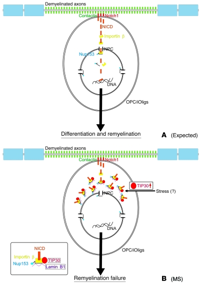
Oligodendrocyte precursor cells (OPCs) persist near the demyelinated axons arising in MS but inefficiently differentiate into oligodendrocytes and remyelinate these axons. The pathogenesis of differentiation failure remains elusive. We initially hypothesized that injured axons fail to present Contactin, a positive ligand for the oligodendroglial Notch1 receptor to induce myelination, and thus tracked axoglial Contactin/Notch1 signaling in situ, using immunohistochemistry in brain tissue from MS patients containing chronic demyelinated lesions. Instead, we found that Contactin was saturated on demyelinated axons, Notch1-positive OPCs accumulated in Contactin-positive lesions, and the receptor was engaged, as demonstrated by cleavage to Notch1-intracellular domain (NICD). However, nuclear translocalization of NICD, required for myelinogenesis, was virtually absent in these cells. NICD and related proteins carrying nuclear localization signals were associated with the nuclear transporter Importin but were trapped in the cytoplasm. Abnormal expression of TIP30, a direct inhibitor of Importin, was observed in these OPCs. Overexpression of TIP30 in a rat OPC cell line resulted in cytoplasmic entrapment of NICD and arrest of differentiation upon stimulation with Contactin-Fc. Our results suggest that extracellular inhibitory factors as well as an intrinsic nucleocytoplasmic transport blockade within OPCs may be involved in the pathogenesis of remyelination failure in MS.


Figures

Figure 1
Figure 1. Remyelination failure in MS may not be due to loss of axonal Contactin stimuli or induction of nuclear Hes5 by astrocytes.
(A) LFB staining showing normal-appearing white matter (blue) and a demyelinated lesion (white) in an MS brain (frontal lobe cerebral lesion from 47-year-old female with secondary progressive MS; disease duration: 20 years). Cntn, Contactin. (B) Section consecutive to A, showing that the expression of Contactin is upregulated in the demyelinated lesion. (C) Demyelination is not apparent in a control subject (parietal lobe cerebral specimen from 81-year-old female without brain disease). (D) Section consecutive to C showing that Contactin is expressed at a low degree; thus, the expression is unremarkable in photograph at lower magnification. Overexpression of Contactin in MS demyelinated lesions are observed in all 10 MS specimens included in this study (the other 9 MS samples are shown in Supplemental Figure 1). (E and F) Contactin immunoreactivity was lost in demyelinated areas where severe tissue damage was evident (asterisks; frontal lobe cerebral lesion from 59-year-old male with primary progressive MS. Isotype controls are shown in Supplemental Figure 2. (GI) Overexpression of Contactin in MS demyelinated lesions occurs on the surfaces of axons positive for neurofilament (NF). Photographs at lower magnification are shown in Supplemental Figure 3. (JL) Preserved OPCs in MS lesions are positive for nuclear Olig1 (24) and express Hes-5 in the cytoplasm but not in the nucleus (arrows and insets). Scale bars: 500 μm (AF); 50 μm (GL); 10 μm (insets in JL).
Figure 2
Figure 2. Accumulations of Notch1-positive OPCs and receptor activation take place; nonetheless, nuclear translocalization of NICD fails in OPCs in MS lesions.
(AC) Notch1-positive cells are increased in a Contactin-positive demyelinated MS lesion in comparison with normal-appearing white matter (border demarcated by broken line). (DF) Immunostaining in a section consecutive to AC with a specific Ab against NICD, which does not recognize full-length Notch1, reveals that the activation of Notch1 and consequent cleavage to NICD took place in these cells. (G) LFB staining in a section consecutive to DF is shown. (H) A photograph at higher magnification showing that NICD, which is expected to translocate into nucleus, is aggregated in cytoplasm but not nucleus (arrows) in the OPCs (see also Supplemental Figure 5 for results from other MS specimens). (IK) These NICD-positive cells are positive for nuclear Olig1, indicative of oligodendroglial cells recruited for remyelination (24) (arrows). DE, demyelinated lesion; NAWM, normal-appearing white matter; v, vessels. Scale bars: 100 μm (AG); 40 μm (H); 50 μm (IK).
Figure 3
Figure 3. Nuclear transporter Importin β and NPC protein Nup153 are not diminished but are aggregated with NICD in cytoplasm of OPCs in MS lesions.
(AC) The expression of Importin β, an indispensable nuclear transport protein, is increased in an MS demyelinated lesion and colocalized with the NICD aggregates in the OPCs (arrows and arrowhead in inset). Photographs at lower magnification are shown in Supplemental Figure 6; note that most, if not all, of these cytoplasmic NICD–aggregated cells are positive for Olig1, an OPC marker (24), as shown in Figure 2. (DF) NPC protein Nup153, required for entrance into the nucleus, is also expressed in the OPCs (arrows). (GI) Photographs at higher magnification showing that the expression of Nup153 is localized at the nuclear envelope and also colocalized with the NICD-cytoplasmic aggregates. (JL) NICD aggregates are not localized within ER structures (positive for PDI). N, nucleus; NE, nuclear envelope; C, cytoplasm. Scale bars: 50 μm (AF); 10 μm (insets in C; JL); 5 μm (GI; insets in JL).
Figure 4
Figure 4. Abnormal expression of Lamin B1 and TIP30 in OPCs in MS lesions.
(A) The expression of Lamin B1 is virtually undetectable in normal cerebral white matter from a control subject. (B) The expression is increased in a MS demyelinated lesion (surrounded by broken line) compared with nearby normal-appearing white matter. (CE; higher magnification) Overexpressed Lamin B1 colocalizes with cytoplasmic NICD aggregates in the OPCs (note that the most of these cytoplasmic NICD–aggregated cells are positive for Olig1, an OPC marker [ref. 24], as shown in Figure 2), but not with nuclear envelopes. (F) TIP30 is undetectable except in small vessels in normal cerebral white matter from a control subject. (G) TIP30 expression is markedly increased in a MS demyelinated lesion (demyelinated area and normal-appearing white matter demarcated by broken line). (HJ) Overexpressed TIP30, an inhibitor of Importin β (19), is associated with Importin β in the OPCs (arrows). (KM; higher magnification) In a few cells with relatively lower expression of TIP30 (e.g., the lower cell in the pictures), Importin β that is not colocalized with TIP30 (arrows) gathers near the nuclear envelope (arrowheads); some appears to have entered the nucleus. Scale bars: 250 μm (A, B, F, and G); 10 μm (CE and KM); 25 μm (HJ).
Figure 5
Figure 5. TIP30-positive cells are substantially increased in chronically demyelinated plaques.
(A and B) Two consecutive sections were chosen for each MS case, and 1 was stained with LFB plus NFR to identify chronically demyelinated plaques and remyelinating shadow plaques (demarcated by broken line), while the other was double-labeled against NICD and TIP30 (occipital lobe cerebral lesion from 51-year-old female with secondary progressive MS is shown). (C) A photograph at higher magnification from a remyelinating shadow plaque in B showing nuclear NICD–positive cells (arrows; the majority of these cells were positive for nuclear Olig1, an OPC marker [ref. 24] as shown in inset). (D) A photograph at higher magnification from a chronically demyelinated plaque in B, showing cytoplasmic TIP30 (cytoplasmic NICD–aggregated) cells (arrowheads). (E and F) Graphs showing results of statistical analysis for the densities of nuclear NICD–positive cells and cytoplasmic TIP30–positive (cytoplasmic NICD–aggregated) cells, respectively. Data representing the same specimens are connected with lines. Bold lines indicate the average of results from 9 specimens. Circles indicate relapse-remitting MS, triangles indicate secondary-progressive MS, and X’s indicate primary-progressive MS. The difference in the density of nuclear NICD–positive cells between chronically demyelinated plaques and remyelinating shadow plaques was not statistically significant (P = 0.069; note that nuclear NICD is rapidly turned over and therefore difficult to detect; see main text), whereas a significant increase in the density of TIP30-positive cells was noted in chronically demyelinated plaques when compared with remyelinating shadow plaques (P = 0.024). Scale bars: 200 μm (A and B); 50 μm (C and D); 20 μm (insets in C).
Figure 6
Figure 6. TIP30 forms cytoplasmic aggregates upon Contactin stimulation and increases susceptibility to cell death in vitro.
(AE) A series of captured pictures from a time-lapse recording of EGFP-TIP30–transfected OLN-93 cells upon Contactin stimulation in vitro. TIP30, initially expressed diffusely in the cytoplasm, becomes confined to perinuclear areas within a few hours after Contactin stimulation. The original recording is available as Supplemental Video 1. (F and G) Photographs at higher magnification of prestimulation (0 minutes) and post-Contactin stimulation (240 minutes) from AE, showing perinuclear cytoplasmic aggregations of TIP30 upon Contactin stimulation. (HM) In those cells with relatively high expressions of TIP30, perinuclear aggregations of TIP30 upon Contactin stimulation resulted in breakdown of the nuclear envelope and subsequent cell death (as seen in M) within several hours. The original recording is available as Supplemental Video 2 (nontransgenic control is shown in Supplemental Video 3). Scale bars: 40 μm (AE); 20 μm (FM).
Figure 7
Figure 7. Inhibition of nuclear translocalization of NICD and myelin protein expressions by TIP30 in vitro.
(A and B) When OLN-93 cells are induced to differentiate via Contactin stimulation for 29 hours, nuclear translocalization of NICD occurs in naive cells but not in TIP30-positive cells (arrows). NICD in such cells are trapped in cytoplasm (arrowheads in inset). (C) TIP30 is expressed in the cytoplasm, outside the ER (the region positive for PDI), similarly to OPCs in MS lesions. (D) Nuclear translocation ratio of NICD is reduced about 60% (from 34% to 14%) by TIP30. Note that the result is likely to be an underestimate because of cell death induced by TIP30 overexpression, as shown in Figure 6. (EH) Contactin stimulation, in contrast to BSA control, promotes the expression of MAG in naive OLN-93 cells (12) but not in TIP30-positive cells (asterisks). Similar results were obtained for CNPase (Supplemental Figure 7). Note that pictures shown are 1D-rendered images from Z stack series (confocal slice image is shown in Figure 6). Error bars in D denote SD. Scale bars: 50 μm (A and B); 20 μm (inset in A); 10 μm (C); 50 μm (EH).
Figure 8
Figure 8. TIP30 inhibits morphological differentiation of OPC cells in vitro.
When naive OLN-93 cells are induced to differentiate via Contactin stimulation for 29 hours, morphological differentiation (an increase in the percentage of differentiated webbed cells [P = 0.001] and decreases in the percentages of cell without processes [P < 0.001] and mono-/bipolar cells [P = 0.005]) was observed in comparison with BSA control. These changes were not observed (statistically not significant; P > 0.05) in TIP30-positive OLN-93 cells, suggesting TIP30 inhibits the morphological differentiation of OPC cells. Morphology of the cells was categorized into 5 groups (see Methods), and their typical appearances are shown. Error bars denote SD. Scale bars: 10 μm.
Figure 9
Figure 9. TIP30 inhibits oligodendroglial nucleocytoplasmic transport and arrests remyelination in MS.
(A) A schematic drawing to show what is theoretically expected during remyelination when the preserved OPCs are naive. Oligodendroglial Notch1 is cleaved to NICD upon its ligation with axonal Contactin. NICD is then translocated into nucleus by nuclear transporter Importin β in an NLS-dependent manner, promoting the expression of myelin-related genes and thereby inducing the maturation of oligodendrocytes for remyelination. (B) Conclusion of the present study of chronically demyelinated plaques in MS. Even though NICD is formed in the preserved OPCs, its nuclear translocalization is inhibited by TIP30, which directly blocks Importin β, thereby causing remyelination failure. TIP30 inhibits both the expression of myelin proteins and morphological differentiation. The abnormal expression of TIP30 may be mediated by extracellular stresses (see Discussion). iOligs, immature oligodendrocytes.

Comment in

Similar articles

Cited by

References

    1. Patrikios P., et al. Remyelination is extensive in a subset of multiple sclerosis patients. Brain. 2006;129:3165–3172. doi: 10.1093/brain/awl217. - DOI - PubMed
    1. Patani R., Balaratnam M., Vora A., Reynolds A. Remyelination can be extensive in multiple sclerosis despite a long disease course. Neuropathol. Appl. Neurobiol. 2007;33:277–287. doi: 10.1111/j.1365-2990.2007.00805.x. - DOI - PubMed
    1. Wolswijk G. Chronic stage multiple sclerosis lesions contain a relatively quiescent population of oligodendrocyte precursor cells. J. Neurosci. 1998;18:601–609. - PMC - PubMed
    1. Chang A., Tourtellotte W.W., Rudick R., Trapp B.D. Premyelinating oligodendrocytes in chronic lesions of multiple sclerosis. N. Engl. J. Med. 2002;346:165–173. doi: 10.1056/NEJMoa010994. - DOI - PubMed
    1. Franklin R.J. Why does remyelination fail in multiple sclerosis? Nat. Rev. Neurosci. 2002;3:705–714. doi: 10.1038/nrn917. - DOI - PubMed

Publication types

MeSH terms