. 2009 Feb;69(2-3):174-90.
doi: 10.1002/dneu.20690.

The JIP3 scaffold protein UNC-16 regulates RAB-5 dependent membrane trafficking at C. elegans synapses

Affiliations

The JIP3 scaffold protein UNC-16 regulates RAB-5 dependent membrane trafficking at C. elegans synapses

Heather M Brown et al. Dev Neurobiol. 2009 Feb.

Abstract

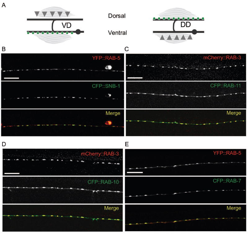
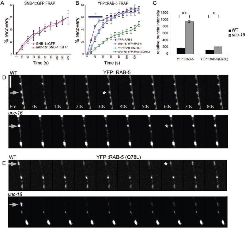
How endosomes contribute to the maintenance of vesicular structures at presynaptic terminals remains controversial and poorly understood. Here, we have investigated synaptic endosomal compartments in the presynaptic terminals of C. elegans GABAergic motor neurons. Using RAB reporters, we find that several subsynaptic compartments reside in, or near, presynaptic regions. Loss of function in the C. elegans JIP3 protein, UNC-16, causes a RAB-5-containing compartment to accumulate abnormally at presynaptic terminals. Ultrastructural analysis shows that synapses in unc-16 mutants contain reduced number of synaptic vesicles, accompanied by an increase in the size and number of cisternae. FRAP analysis revealed a slow recovery of RAB-5 in unc-16 mutants, suggestive of an impairment of RAB-5 activity state and local vesicular trafficking. Overexpression of RAB-5:GDP partially suppresses, whereas overexpression of RAB-5:GTP enhances, the synaptic defects of unc-16 mutants. Our data demonstrate a novel function of UNC-16 in the regulation of synaptic membrane trafficking and suggest that the synaptic RAB-5 compartment contributes to synaptic vesicle biogenesis or maintenance.


Figures

Figure 1
Figure 1. Endosomal markers localize to synapses
(A) GABAergic DD and VD neurons are pseudo-unipolar such that their cell bodies (gray dots) are on the ventral side of the animal and synapses (green circles) form en passant on the dorsal process of the DD neurons and the ventral process of the VD neurons (gray lines). Synaptic inputs (gray triangles) are made onto the ventral processes of the DD or the dorsal processes of the VD neurons (B-E) Confocal images of animals co-expressing XFP∷RAB, or SNB-1∷CFP transgenes. (B) Punc-25-YFP∷RAB-5 (red) and Punc-25-SNB-1∷CFP (green) expression in the ventral process of VD neurons. (C) Punc-25-mCherry∷RAB-3 (red) and Punc-25-CFP∷RAB-11 (green) expression in the dorsal process of the DD neurons. (D) Punc-25-mCherry∷RAB-3 (red) and Punc-25-CFP∷RAB-10 (green) expression in the dorsal process of the DD neurons. (E) Punc-25-YFP∷RAB-5 (red) and Punc-25-CFP∷RAB-7 (green) expression in the dorsal process of the DD neurons. Scale bars are 10μm.
Figure 2
Figure 2. Endosomal compartments are differentially regulated by kinesin-1 and UNC-104 motor proteins
Images are confocal z-stack projections of WT, unc-104 and unc-116 mutant animals showing expression of (A) YFP∷RAB-5, (B) CFP∷RAB-7, (C) CFP∷RAB-10 and (D) CFP∷RAB-11. In unc-116 mutants, RAB-5 and RAB-7 puncta intensity and size decrease. In unc-104 mutants, RAB-5 becomes entirely diffuse, puncta are rarely detectable; RAB-7 shows similar, but weaker, defects. Quantitation of XFP∷RAB puncta per 100 μm is shown in (E) as mean ± SEM(*P< 0.05, **P< 0.01 compared to WT, n = 8-10 animals for each genotype, n/s = not significant). Scale bars are 10 μm.
Figure 3
Figure 3. Loss of function in unc-16 specifically alters synaptic endosomal YFP∷RAB-5
(A) Images are confocal z-stack projections of YFP∷RAB-5 in the dorsal cords of WT and mutants as labeled. In order to show the full dynamic range of the YFP∷RAB-5 puncta fluorescence all images for unc-16 containing animals were taken with reduced laser power (see Methods). In WT, YFP∷RAB-5 is localized along the nerve processes in a punctate distribution. In unc-16 mutants puncta size and intensity are increased, and the punctate pattern along the nerve process becomes irregular in size and distribution. In dhc-1 mutant animals, YFP∷RAB-5 fluorescence becomes more dispersed with decreased puncta size and intensity. unc-16 unc-116, unc-16; unc-104, and unc-16;dhc-1 double mutants show an additive phenotype, with YFP∷RAB-5 puncta seen as increased intensity but variable size. (B) Quantitation of YFP∷RAB-5 puncta area and intensity. For puncta area (μm2), data is depicted as average puncta area (bars) and individual puncta area (black circles). For puncta intensity, RIU refers to Relative Intensity Units of peak puncta intensities. All data are shown as mean ± SEM, statistical comparison is made pairwise with WT (red lines) or pairwise with each other (black lines) (n=10 animals for each genotype, *P< 0.05 and **P<0.01). (C-D) CFP∷RAB-7 is largely unaltered in unc-16 mutants. (C). Images are confocal z-stack projections of CFP∷RAB-7 in the dorsal cords of WT and unc-16(ju146). (D) Quantitation of CFP∷RAB-7 puncta size and intensity are shown as mean ± SEM (*P< 0.05 compared to WT, n=10 animals for each genotype, n/s = not significant). Scale bars are 10μm.
Figure 4
Figure 4. The effect of unc-16 on the RAB-5 compartment is distinct from those in mutants defective in synaptic vesicle endocytosis
(A) Confocal z-stack projections of YFP∷RAB-5 in the dorsal cords of AP180/unc-11(e47), endophilin/unc-57(e406), synaptojanin/unc-26(e205), unc-16; unc-11, unc-16; unc-57, and unc-16; unc-26. In unc-11 mutants YFP∷RAB-5 puncta size and intensity are decreased, while in unc-57 and unc-26 mutants YFP∷RAB-5 puncta are dispersed and diffuse. The double mutants with unc-16 show an additive phenotype with increased intensity. In order to show the full dynamic range of the YFP∷RAB-5 puncta fluorescence all images for unc-16 containing animals were taken with reduced laser power (see Methods) (B) Quantitation of puncta area and intensity. Data presentation is the same as in Fig. 2 B; red lines, pairwise comparison with WT, black lines, pairwise comparison with each other (n=10 animals for each genotype, *P< 0.05 and **P<0.01). (C) Confocal z-stack projections of SNB-1∷GFP in the dorsal cords of WT, unc-16, unc-57 and unc-16; unc-57 mutants. In unc-57 mutants SNB-1∷GFP puncta become dispersed with few distinct puncta remaining. unc-16; unc-57 double mutants show an intermediate phenotype with puncta number more closely resembling that of unc-16 single mutants, however, puncta area and distribution are irregular compared with either single mutant alone. (D) Quantitation of SNB-1∷GFP puncta number per 100 μm in unc-16, unc-57 and the double mutant (mean ± SEM, *P< 0.05, n=7-10 animals for each genotype); red lines, pairwise comparison with WT; black lines, pairwise comparison with each other. Scale bars are 10μm.
Figure 5
Figure 5. Vesicular structures at the synapses of unc-16 mutant animals are altered
(A-D) SNB-1∷GFP, not mCherry∷RAB-3, puncta are altered in unc-16 mutants. (A) Images are confocal z-stack projections of SNB-1∷GFP in the dorsal cords of WT, unc-16(ju146) and unc-16(e109) mutant animals. (B) Images are single-plane confocal scan of YFP∷RAB-5 and SNB-1∷CFP co-expression in unc-16 mutants, showing that these markers are affected similarly. (C) mCherry∷RAB-3 puncta are unaltered in unc-16(ju146) animals. Images are confocal z-stack projections of the dorsal cord in young adults. (D) Quantitation of SNB-1∷GFP and mCherry∷RAB-3 in WT and unc-16 mutant animals (puncta area μm2, mean ± SEM, *P< 0.05 compared to WT). Scale bars are 10μm. (E) Shown are TEM images of ventral cord GABAergic synapses of WT and two examples of unc-16. Overall synaptic morphology is normal but vesicular structures are altered in unc-16(ju146) mutant animals. Dense core vesicles (dcv), synaptic vesicles (sv), the active zone (AZ) and cisternae (c) are labeled with black arrows. Abnormal cisternal structures are labeled with a red arrow. Scale bar is 0.5 μm. (F) Quantitation of the number of SV per synapse and of total cisternae per 45 nm section in WT, unc-16(ju146) and unc-16(e109) (mean ± SEM, GABAergic: WT n= 21, unc-16(ju146) n= 6 and unc-16(e109) n= 10; Cholinergic: WT n= 46, unc-16(ju146) n= 14 and unc-16(e109) n= 14)
Figure 6
Figure 6. Loss of unc-16 function impairs RAB-5 recovery rates after photobleaching
(A) Averaged FRAP recovery profiles of SNB-1∷GFP (juls1) in WT (diamonds) and unc-16 (squares) (mean ± SEM, n=5). Recovery rates of SNB-1∷GFP are unaltered in unc-16 mutants. (B) Averaged FRAP recovery profiles of YFP∷RAB-5 (juls198) in WT (diamonds) and unc-16 (squares) and of YFP∷RAB-5(Q78L) in WT (circles) and unc-16 (triangles) ((mean ± SEM, * p< 0.01, n=5). Recovery rates of YFP∷RAB-5 after photobleaching are significantly slower in unc-16 mutants than in WT animals. Recovery rates of YFP∷RAB-5(Q78L) in WT are similar to those of YFP∷RAB-5 in unc-16 mutants. YFP∷RAB-5(Q78L) is slowed further in unc-16 mutants. (C) Quantitation of YFP∷RAB-5 and YFP∷RAB-5(Q78L) puncta intensity in WT and unc-16 mutants are shown as mean ± SEM.) (n=10 animals for each genotype, *P< 0.05). (D-E) Representative time course is shown for YFP∷RAB-5 recovery in WT and unc-16 mutant, and YFP∷RAB-5(Q78L) in WT and unc-16 mutant. Gray arrows indicate bleached puncta, asterix shows time point where recovery has reached 55%. Scale bar is 5μm.
Figure 7
Figure 7. RAB-5:GDP suppresses and RAB-5:GTP enhances the SNB-1∷GFP phenotype in unc-16
(A). Shown are dorsal cord epifluorescence images of SNB-1∷GFP in WT and unc-16 mutant animals. unc-16 animals expressing RAB-5(S33N) show an improved SNB-1∷GFP pattern; and unc-16 animals expressing RAB-5(Q78L) show disorganization and aggregation of the SNB-1∷GFP pattern. Scale bar is 10μm. (B) Quantitation of SNB-1∷GFP phenotypes The morphology of SNB-1∷GFP puncta was categorized as normal (~0.86 μm2), enlarged (~1.1 μm2) or aggregate (observed as a stretch of irregularly distributed puncta with varying size up to 3.5 μm2) * p< 0.01, n= 34-49 animals per genotype.

References

    1. Bonanomi D, Benfenati F, Valtorta F. Protein sorting in the synaptic vesicle life cycle. Prog Neurobiol. 2006;80:177–217. - PubMed
    1. Cavalli V, Kujala P, Klumperman J, Goldstein LS. Sunday Driver links axonal transport to damage signaling. J Cell Biol. 2005;168:775–787. - PMC - PubMed
    1. Dittman JS, Kaplan JM. Factors regulating the abundance and localization of synaptobrevin in the plasma membrane. Proc Natl Acad Sci U S A. 2006;103:11399–11404. - PMC - PubMed
    1. Hallam SJ, Jin Y. lin-14 regulates the timing of synaptic remodelling in Caenorhabditis elegans. Nature. 1998;395:78–82. - PubMed
    1. Hamill DR, Severson AF, Carter JC, Bowerman B. Centrosome maturation and mitotic spindle assembly in C. elegans require SPD-5, a protein with multiple coiled-coil domains. Dev Cell. 2002;3:673–684. - PubMed

Publication types

MeSH terms

Substances