. 2009 Feb 27;284(9):5742-52.
doi: 10.1074/jbc.M808507200. Epub 2008 Dec 23.

Deubiquitination of CXCR4 by USP14 is critical for both CXCL12-induced CXCR4 degradation and chemotaxis but not ERK ativation

Affiliations

Deubiquitination of CXCR4 by USP14 is critical for both CXCL12-induced CXCR4 degradation and chemotaxis but not ERK ativation

Marjelo A Mines et al. J Biol Chem. .

Abstract

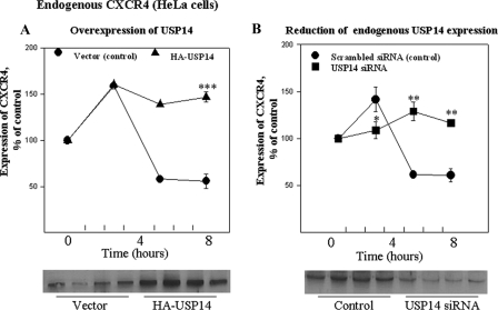
The chemokine receptor CXCR4 plays important roles in the immune and nervous systems. Abnormal expression of CXCR4 contributes to cancer and inflammatory and neurodegenerative disorders. Although ligand-dependent CXCR4 ubiquitination is known to accelerate CXCR4 degradation, little is known about counter mechanisms for receptor deubiquitination. CXCL12, a CXCR4 agonist, induces a time-dependent association of USP14 with CXCR4, or its C terminus, that is not mimicked by USP2A, USP4, or USP7, other members of the deubiquitination catalytic family. Co-localization of CXCR4 and USP14 also is time-dependent following CXCL12 stimulation. The physical interaction of CXCR4 and USP14 is paralleled by USP14-catalyzed deubiquitination of the receptor; knockdown of endogenous USP14 by RNA interference (RNAi) blocks CXCR4 deubiquitination, whereas overexpression of USP14 promotes CXCR4 deubiquitination. We also observed that ubiquitination of CXCR4 facilitated receptor degradation, whereas overexpression of USP14 or RNAi-induced knockdown of USP14 blocked CXCL12-mediated CXCR4 degradation. Most interestingly, CXCR4-mediated chemotactic cell migration was blocked by either overexpression or RNAi-mediated knockdown of USP14, implying that a CXCR4-ubiquitin cycle on the receptor, rather than a particular ubiquitinated state of the receptor, is critical for the ligand gradient sensing and directed motility required for chemokine-mediated chemotaxis. Our observation that a mutant of CXCR4, HA-3K/R CXCR4, which cannot be ubiquitinated and does not mediate a chemotactic response to CXCL12, indicates the importance of this covalent modification not only in marking receptors for degradation but also for permitting CXCR4-mediated signaling. Finally, the indistinguishable activation of ERK by wild typeor 3K/R-CXCR4 suggests that chemotaxis in response to CXCL12 may be independent of the ERK cascade.


Figures

FIGURE 1.
FIGURE 1.
CXCR4 selectively interacts with USP14 via the C terminus of the receptor. HEK293 cells stably expressing Myc-CXCR4 were exposed to CXCL12 (10 nm) for the indicated time intervals (A-C). Myc-CXCR4 was immunoprecipitated (IP) from cell lysates using a mouse anti-Myc antibody (see “Experimental Procedures”). A, CXCL12 causes a time-dependent association of CXCR14 with USP14. The amount of co-precipitated USP14 protein was detected by Western blotting for the HA epitope on USP14. The membrane was stripped and reprobed using a rabbit anti-Myc antibody to evaluate Myc-CXCR4 loading. The migration of molecular weight markers is shown to the left of the gel. Data shown are representative of one experiment performed six times. IB, immunoblot. B, quantitation of the relative amount of USP14 co-precipitated with CXCR4 was determined by densitometric scanning as outlined under “Experimental Procedures”; n = 6. C, selectivity of USP-isoform interaction with Myc-CXCR4. Experiments were performed as in A and described in detail under “Experimental Procedures.” There was no detectable interaction of the CXCR4 with USP7 (data not shown). Co-precipitated HA-USP2a (▴, n = 4) and His-USP4 (▪, n = 3) were detected using anti-HA and anti-His antibodies (see “Experimental Procedures”). D, USP14 interacts with the C terminus of CXCR4; GSH-Sepharose-bound GST (lane 2, control) or GST-CXCR4 C-terminal fusion protein (lane 3) was incubated with HEK293 cell lysates prepared from control (i.e. not stimulated by CXCL12 ligand), as described under “Experimental Procedures.” Upper panel, HA-USP14 was detected by Western blotting using an anti-HA antibody. Lower panel, GST was detected using a rabbit anti-GST antibody. An aliquot of the cell lysate is shown in lane 1. Data shown in B and C are mean ± S.E. from the number of independent experiments outlined above. *, p < .05; **, p < .01; ***, p < .001, compared with cells not stimulated with CXCL12 (control).
FIGURE 2.
FIGURE 2.
CXCL12 enhances apparent USP14 co-localization with CXCR4. HEK293 cells stably expressing EGFP-CXCR4 and transiently transfected with HA-USP14 were treated without (control) or with CXCL12 (10 nm) for the indicated time intervals. The cells were fixed and evaluated using immunohistochemistry via confocal microscopy as detailed under “Experimental Procedures.” Representative laser-scanning confocal micrographs demonstrating the distribution of EGFP-CXCR4 (green), USP14 (red), and overlay (yellow) are shown. Images were processed using Photoshop software.
FIGURE 3.
FIGURE 3.
USP14 modulates CXCR4 ubiquitination. A, antibody directed against endogenous ubiquitin reveals the time-dependent ubiquitination of Myc-CXCR4 (upper panel) in response to CXCL12 (10 nm) treatment of HEK293 cells (see “Experimental Procedures”). The lower panel, obtained by reprobing the Western blot with an anti-Myc antibody directed against the Myc-CXCR4, readily detects the receptor protein migrating at ∼45,000, but not the higher molecular mass “ladders” at 54 kDa and above. IP, immunoprecipitation; IB, immunoblot. B, quantitation of the relative density of bands representing CXCR4-endogenous Ub complexes was determined by densitometric scanning (•; see “Experimental Procedures”). Superimposed on these data are the findings from the time course of USP14 association with the CXCR4 in CXCL12-exposed cells (○), reported in Fig. 1B, for comparison. C, overexpression of USP14 eliminates detectable CXCR4 ubiquitination in response to CXCL12. HEK293 cells stably expressing Myc-CXCR4 and transiently transfected with vector alone (Vector) or HA-USP14 were incubated for 10 min with CXCL12 as in A. This incubation was terminated immediately for some samples (10 min, no recovery) but allowed to continue after washing away the CXCL12, for 60 min (10 min, +60 min recovery). The gel data shown below the bar graph, from one representative experiment, confirm that transfection of the HEK293 cells with the cDNA encoding HA-USP14 indeed leads to overexpression of this enzyme. D, RNA interference knockdown of endogenous USP14 eliminates deubiquitination of CXCL12-evoked ubiquitination of Myc-CXCR4. HEK293 cells transfected with scrambled (control) siRNA or USP14 siRNAs were treated without CXCL12 (none) or with 10 nm CXCL12 for 10 min (10 min, no recovery) or 10 min followed by a 60-min recovery period (10 min, + 60 min recovery) as described under “Experimental Procedures.” The panel below the bar graph provides a representative gel that confirms the ability of the siRNA construct to successfully reduce the expression of the USP14 protein in these cells under these conditions. Myc-CXCR4 was isolated by immunoprecipitation with an anti-Myc antibody, and the CXCR4-Ub complexes were quantified by Western blot using an antibody against endogenous ubiquitin. Blots were stripped and reprobed using a Myc antibody to evaluate loading of Myc-CXCR4. Data in B-D represent the mean ± S.E. from three independent experiments. *, p < 0.05; **, p < 0.01; ***, p < 0.001, compared with controls cells (no CXCL12 treatment).
FIGURE 4.
FIGURE 4.
USP14 prevents CXCL12-mediated EGFP-CXCR4 degradation and increases the steady state level of the receptor. A, HEK293 cells stably expressing EGFP-CXCR4 were treated with CXCL12 for 8 h (+); this prolonged incubation allowed detection of CXCL12-evoked receptor down-regulation. EGFP-CXCR4 levels were detected by Western blot using an anti-EGFP antibody. B, quantitation of the relative amount of CXCR4 was determined by densitometric scanning as outlined under “Experimental Procedures.” Data are mean ± S.E. from three independent experiments. *, p < 0.05; compared with control cells (no CXCL12 treatment (-)). ns, not statistically significant.
FIGURE 5.
FIGURE 5.
Degradation of endogenous CXCR4 in HeLa cells is disrupted by modulation of USP14 expression. A, HeLa cells transiently transfected with vector or HA-USP14 were treated with CXCL12 for 0, 3, 5, or 8 h as indicated. CXCR4 levels were detected by Western blot using an anti-CXCR4 antibody. Quantitation of relative amount of CXCR4 was determined by densitometric scanning as outlined under “Experimental Procedures.” Data obtained at time 0 and 8 h Data are mean ± S.E. from three independent experiments. *, p < 0.05; **, p < 0.01; ***, p < 0.001 compared with control cells (no CXCL12 treatment, i.e. time 0); the data obtained at 3 and 5 h after CXCL12 incubation are from only two independent experiments, and thus statistical analyses were not performed for these time points. B, HeLa cells transiently transfected with scrambled siRNA (control) or USP14-specific siRNA were treated with CXCL12 for 0, 3, 5, or 8 h, as indicated. CXCR4 levels were detected by Western blot using an anti-CXCR4 antibody. Quantitation of relative amount of CXCR4 was determined by densitometric scanning as outlined under “Experimental Procedures.” Data are mean ± S.E. from three independent experiments. *, p < 0.05; **, p < 0.01.
FIGURE 6.
FIGURE 6.
CXCR4-Ub cycle is essential for CXCR4-mediated chemotaxis. A, chemotaxis was evaluated in HEK293 cells stably expressing Myc-CXCR4 (and endogenous levels of USP14, i.e. “vector alone”) or in cells overexpressing HA-USP14 as described in detail under “Experimental Procedures.” Overexpression of USP14, which dramatically reduces CXCL12-evoked CXCR4 ubiquitination (cf. Fig. 3C), also dramatically attenuates CXCL12-evoked chemotaxis. B, knockdown of endogenous USP14 expression with USP14-directed siRNA, which leads to enhanced CXCL12-induced CXCR4 ubiquitination (cf. Fig. 3D), also significantly reduces CXCL12-induced chemotaxis. C, chemotaxis in response to CXCL12 was evaluated in cells expressing an HA-WT CXCR4 and in cells expressing a 3K/R mutant receptor, as described under “Experimental Procedures.” Taken together, A and B suggest that the CXCR4-Ub cycle, and not a particular ubiquitinated state of CXCR4, is essential for CXCL12-mediated chemotaxis, and the data in C confirm that it is the CXCR4 molecule itself that must undergo a ubiquitination/deubiquitination cycle for chemotaxis to occur. Values represent the mean ± S.E. from three independent experiments performed in duplicate. All chemotaxis data are expressed as the chemotactic index, which is calculated as the ratio of the number of cells that migrate across the Boyden chamber in the presence of CXCL12 at a given concentration compared with the number of cells migrating in the absence of CXCL12; the value of 1 means that there was no migration greater than that observed in control, nonstimulated cells. Data were analyzed using Student's unpaired t test. *, p < 0.05; **, p < 0.01; ***, p < 0.001, and compared with control cells, e.g. cells expressing endogenous USP14 (A), scrambled siRNA (B), or HA-WT CXCR4 (C).
FIGURE 7.
FIGURE 7.
ERK activation by CXCR4 occurs independently of the ability of the CXCR4 to be ubiquitinated. A, ERK activation was evaluated in HEK293 cells transiently transfected with HA-WT CXCR4 or HA-3K/R CXCR4 and stimulated with CXCL12 (10 nm) for 0, 5, 15, 30, or 60 min, and ERK activity in cell lysates assessed was as described under “Experimental Procedures.” B, quantitation of ERK activation was based on the amount of ERK detected using an anti-P-ERK antibody; total ERK, assessed using the ERK2 antibody, was indistinguishable in all conditions, and thus the data were not normalized to total ERK. Data are mean ± S.E. from three independent experiments.

References

    1. Haribabu, B., Richardson, R. M., Fisher, I., Sozzani, S., Peiper, S. C., Horuk, R., Ali, H., and Snyderman, R. (1997) J. Biol. Chem. 272 28726-28731 - PubMed
    1. Balabanian, K., Lagane, B., Infantino, S., Chow, K. Y., Harriague, J., Moepps, B., Arenzana-Seisdedos, F., Thelen, M., and Bachelerie, F. (2005) J. Biol. Chem. 280 35760-35766 - PubMed
    1. Nagasawa, T., Hirota, S., Tachibana, K., Takakura, N., Nishikawa, S., Kitamura, Y., Yoshida, N., Kikutani, H., and Kishimoto, T. (1996) Nature 382 635-638 - PubMed
    1. Aiuti, A., Webb, I. J., Bleul, C., Springer, T., and Gutierrez-Ramos, J. C. (1997) J. Exp. Med. 185 111-120 - PMC - PubMed
    1. D'Apuzzo, M., Rolink, A., Loetscher, M., Hoxie, J. A., Clark-Lewis, I., Melchers, F., Baggiolini, M., and Moser, B. (1997) Eur. J. Immunol. 27 1788-1793 - PubMed

Publication types