. 2009 Feb 20;284(8):5229-39.
doi: 10.1074/jbc.M805747200. Epub 2008 Dec 23.

Glycogen synthase kinase-3 regulates the phosphorylation of severe acute respiratory syndrome coronavirus nucleocapsid protein and viral replication

Affiliations

Glycogen synthase kinase-3 regulates the phosphorylation of severe acute respiratory syndrome coronavirus nucleocapsid protein and viral replication

Chia-Hsin Wu et al. J Biol Chem. .

Abstract

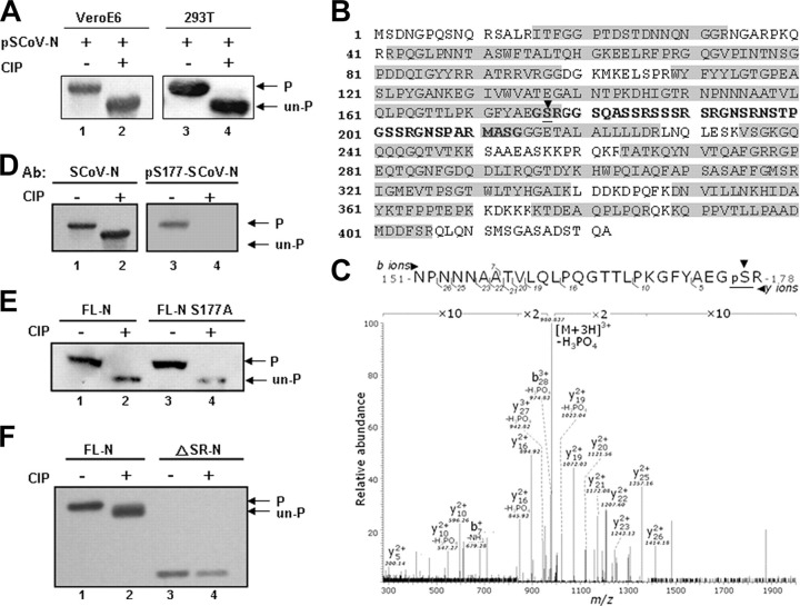
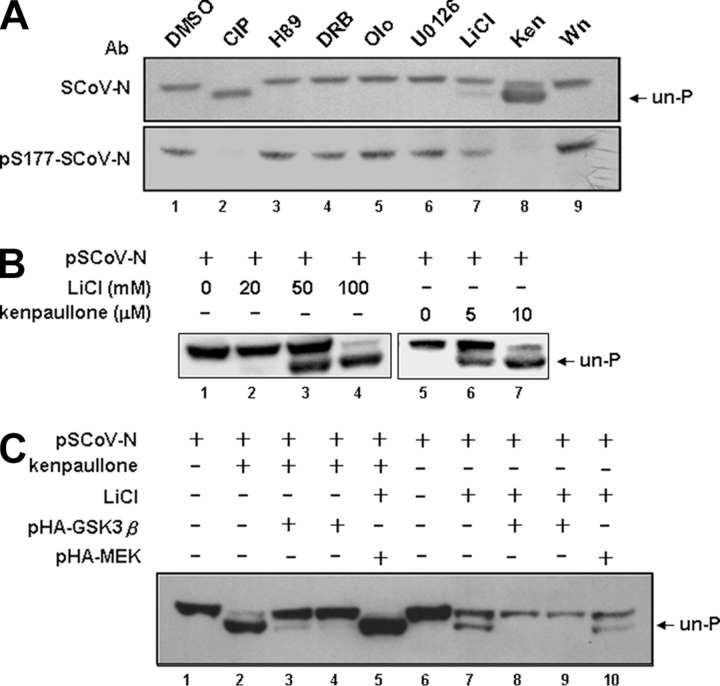
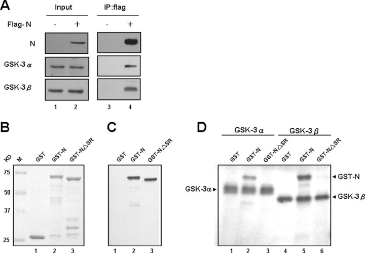
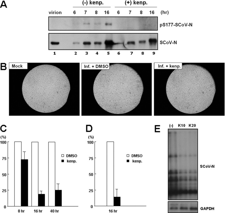
Coronavirus (CoV) nucleocapsid (N) protein is a highly phosphorylated protein required for viral replication, but whether its phosphorylation and the related kinases are involved in the viral life cycle is unknown. We found the severe acute respiratory syndrome CoV N protein to be an appropriate system to address this issue. Using high resolution PAGE analysis, this protein could be separated into phosphorylated and unphosphorylated isoforms. Mass spectrometric analysis and deletion mapping showed that the major phosphorylation sites were located at the central serine-arginine (SR)-rich motif that contains several glycogen synthase kinase (GSK)-3 substrate consensus sequences. GSK-3-specific inhibitor treatment dephosphorylated the N protein, and this could be recovered by the constitutively active GSK-3 kinase. Immunoprecipitation brought down both N and GSK-3 proteins in the same complex, and the N protein could be phosphorylated directly at its SR-rich motif by GSK-3 using an in vitro kinase assay. Mutation of the two priming sites critical for GSK-3 phosphorylation in the SR-rich motif abolished N protein phosphorylation. Finally, GSK-3 inhibitor was found to reduce N phosphorylation in the severe acute respiratory syndrome CoV-infected VeroE6 cells and decrease the viral titer and cytopathic effects. The effect of GSK-3 inhibitor was reproduced in another coronavirus, the neurotropic JHM strain of mouse hepatitis virus. Our results indicate that GSK-3 is critical for CoV N protein phosphorylation and suggest that it plays a role in regulating the viral life cycle. This study, thus, provides new avenues to further investigate the specific role of N protein phosphorylation in CoV replication.


Figures

FIGURE 1
FIGURE 1
The N protein of SCoV is hyperphosphorylated over the central SR-rich motif.A, the N expression plasmid pSCoV-N was transfected into VeroE6 (lanes 1 and 2) and 293T (lanes 3 and 4) cells. The cell lysates, either untreated (lanes 1 and 3) or treated with CIP enzyme (lanes 2 and 4), were assayed by Western blot analysis (probed with an Ab against N protein). The phosphorylated and unphosphorylated N proteins are indicated. B, the purified FLAG-tagged N protein of SCoV from 293T cells was subjected to mass spectrometric analysis. The sequences recovered by the analysis are marked within gray boxes. The serine residue (Ser-177) identified to be phosphorylated is underlined and identified with an arrowhead. C, the results of mass spectrometric analysis, showing the peptide containing Ser-177 to be ∼100% phosphorylated, marked by underlining and an arrowhead. The lysates from 293T cells, transfected with wild type N protein (D), wild type N and mutant S177A N protein (E), or with wild type N and ΔSR-N protein expression constructs (F) were treated with CIP and processed for Western blot analysis. Except for the right panel of D (probed with an Ab recognizing the phosphorylated Ser-177 N protein), the blots of D-E were probed with an Ab against N protein.
FIGURE 2
FIGURE 2
GSK-3 kinase is involved in the phosphorylation of SCoV N protein.A, by treatment with specific inhibitors against kinases predicted by the NetPhos software (for SR-rich motifs), N protein-expressing 293T cell lysates were processed for Western blot analysis (probed with an Ab against N protein and an Ab against phosphorylated Ser-177 N protein). The unphosphorylated form is indicated. The lysate subjected to CIP treatment was used as the control showing the position of unphosphorylated N protein. Wn, wortmannin; Ken, kenpaullone; olo, olomoucine; DRB, 5,6-dichlorobenzimidazole riboside. B, the dose-dependent effects of two specific inhibitors for GSK-3, LiCl and kenpaullone, on the phosphorylation of N were evaluated, with the position of the unphosphorylated N indicated. C, dephosphorylated N resulting from treatment with LiCl and kenpaullone could be recovered by the constitutively active form of GSK-3β (pHA-GSK-3β, an S9A mutant). The control used pHA-MEK, the constitutively active form of MEK, showing the specificity of recovery. The position of the unphosphorylated N protein is indicated.
FIGURE 3
FIGURE 3
Characterization of the interaction between SCoV N and GSK-3 α and β forms and a direct phosphorylation effect of GSK-3 kinases on N by in vitro kinase assays.A, cell lysates from 293T cells transfected with FLAG-N protein construct were processed for coimmunoprecipitation analysis. Immunoprecipitates (IP) were analyzed by Western blot analysis and probed with Abs for N and GSK-3 (α and β) as indicated. B, the input control of GST-fusion proteins used for the in vitro kinase assay was shown by Coomassie Blue staining. C, the control for GST fusion proteins was done by immunoblotting with an Ab against SCoV N protein. D, the in vitro GSK-3α and GSK-3β kinase assays using the GST fusion proteins as substrates. The positions of autophosphorylated GSK-3 kinases are marked, which can serve as the positive controls for the reaction.
FIGURE 4
FIGURE 4
The amino acid residues Ser-189 and Ser-207 of the SR-rich motif are critical for GSK-3 mediated phosphorylation of the SCoV N protein.A, two series of GSK-3 consensus phosphorylation sites were identified within the SR-rich motif (residues 176-214). B, lysates from 293T cells transfected with wild type or mutant N protein expression constructs (mutations at residues 189 and/or 207) were pretreated with kenpaullone (10 μm) or with DMSO (control) and subjected to high resolution NuPAGE gels and Western blot analysis (probed with an Ab against N protein). Lysates from control cells were treated with CIP as a control to mark the position of the dephosphorylated N protein. C, the five protein lysates from panel B as indicated were processed for two-dimensional gel electrophoresis and Western blot analysis (pI range 7-11; probed with an Ab against SCoV N protein). D, wild type (wt) N (with or without CIP treatment) and mutant N proteins containing S177A, S189A, and/or S207A mutations were subjected to Western blotting analysis probed with Ser(P)-177-N and SCoV-N Abs as indicated. E, the wild type FLAG-N and FLAG-S189A/S207A N proteins were used as substrates for GSK-3β in vitro kinase assay. The expression of protein substrates was shown by Coomassie Blue staining (left panel) and Western blotting analysis (middle panel). The positions of phosphorylated FLAG-N and autophosphorylated GSK-3β are marked (right panel, duplicate experiments for each substrate).
FIGURE 5
FIGURE 5
The effects of the GSK-3 inhibitor, kenpaullone, on N protein phosphorylation and the virology of SCoV in the VeroE6 infectious system.A, the phosphorylation status of N proteins in SCoV virions and in virus-infected (Inf.) cells (treated or untreated with kenpaullone (kenp.)) was evaluated by Western blot analysis (probed with Abs against Ser(P)-177-SCoV-N and SCoV-N Abs as indicated). After pretreatment with 10 μm kenpaullone or with DMSO (control), the cells were infected with virus (multiplicity of infection 1), and cell lysates were harvested at different time points for Western blot analysis. B, the effect of kenpaullone pretreatment on virus-induced CPE was evaluated at 16 h after SCoV infection (multiplicity of infection 1) by microscopy. Moreover, the effect of kenpaullone pretreatment on viral titer was evaluated by quantitative PCR (C) and by TCID50 assay (D), with virus harvested from the medium at time as indicated after virus infection. The viral titers from medium treated with DMSO control was set to a value of 1 and the virus titers from kenpaullone pretreatment medium were normalized to this value. E, the effect of kenpaullone pretreatment (10 and 20 μm) on the synthesis of virus RNA was evaluated by Northern blot analysis (probed with DIG-labeled N or glyceraldehyde-3-phosphate dehydrogenase (GAPDH) probes) with cellular RNA harvested 16 h after infection.
FIGURE 6
FIGURE 6
GSK-3 kinase is also involved in the phosphorylation of JHMV N protein.A, only one series of GSK-3 consensus phosphorylation site was identified in SR-rich motif of JHMV N protein (residues 193-213). The potential kinase(s) of each serine residue within this region predicted by NetPhos program was summarized. PKC, protein kinase C. B, wild type (wt) N (with or without CIP treatment) and mutant N proteins containing S197A, S201A, S205A, and S209A mutations isolated from 293T cells were subjected to Western blotting analysis probed with Ser(P)-197-JHMV-N and JHMV-N Abs as indicated. C, by treatment with specific inhibitors against various kinases, N protein-expressing 293T cell lysates were processed for Western blot analysis (probed with Abs against Ser(P)-197-JHMV-N and JHMV-N proteins as indicated). The lysate subjected to CIP treatment was used as the control showing the specificity of Ser(P)-197-JHMV-N Ab. Wn, wortmannin; Ken, kenpaullone; olo, olomoucine; DRB, 5,6-Dichlorobenzimidazole riboside. D, three protein lysates from panel C were processed for two-dimensional gel electrophoresis (pI range 7-11) and for subsequent immunoblotting analysis (probed with Ab against JHMV-N protein).
FIGURE 7
FIGURE 7
Effects of the GSK-3 inhibitors kenpaullone and LiCl on N protein phosphorylation and on the virology of JHMV in the infectious system.A, the phosphorylation status of N proteins in JHMV virions and in virus-infected cells (treated with DMSO control or various kinase inhibitors as indicated) was evaluated by Western blot analysis (probed with Abs against Ser(P)-197-JHMV-N and JHMV-N Abs). The DBT cells were pretreated with the inhibitors for 1 h before JHMV infection (multiplicity of infection 1), and then the cell lysates were harvested at 16 h post-infection. Wn, wortmannin; Ken, kenpaullone; olo, olomoucine; DRB, 5,6-Dichlorobenzimidazole riboside. The effects of specific kinase inhibitors on virus-induced CPE (by microscopy observation) (B) and viral titer (by quantitative PCR assay) (C) were also evaluated at 16 h post-infection. The viral titers from medium treated with DMSO control was set to a value of 1, and the virus titers from inhibitors pretreatment medium were normalized to this value. D, the effect of kenpaullone pretreatment on viral RNA synthesis was evaluated by Northern blot analysis (probed with DIG-labeled JHMV-N protein probes) with cellular RNA extracted at 12 h after infection (multiplicity of infection 1 and 0.3).

References

    1. Drosten C., Gunther S., Preiser W., van der Werf S., Brodt H.R., Becker S., Rabenau H., Panning M., Kolesnikova L., Fouchier R.A., Berger A., Burguiere A.M., Cinatl J., Eickmann M., Escriou N., Grywna K., Kramme S., Manuguerra J.C., Muller S., Rickerts V., Sturmer M., Vieth S., Klenk H.D., Osterhaus A.D., Schmitz H., Doerr H.W. N. Engl. J. Med. 2003;348:1967–1976. - PubMed
    1. Ksiazek T.G., Erdman D., Goldsmith C.S., Zaki S.R., Peret T., Emery S., Tong S., Urbani C., Comer J.A., Lim W., Rollin P.E., Dowell S.F., Ling A.E., Humphrey C.D., Shieh W.J., Guarner J., Paddock C.D., Rota P., Fields B., DeRisi J., Yang J.Y., Cox N., Hughes J.M., LeDuc J.W., Bellini W.J., Anderson L.J. N. Engl. J. Med. 2003;348:1953–1966. - PubMed
    1. Snijder E.J., Bredenbeek P.J., Dobbe J.C., Thiel V., Ziebuhr J., Poon L.L., Guan Y., Rozanov M., Spaan W.J., Gorbalenya A.E. J. Mol. Biol. 2003;331:991–1004. - PMC - PubMed
    1. Lai M.M., Cavanagh D. Adv. Virus Res. 1997;48:1–100. - PMC - PubMed
    1. Fang X., Ye L., Timani K.A., Li S., Zen Y., Zhao M., Zheng H., Wu Z. J. Biochem. Mol. Biol. 2005;38:381–385. - PubMed

Publication types

MeSH terms

Substances