. 2009 Mar 20;284(12):8054-63.
doi: 10.1074/jbc.M805384200. Epub 2008 Dec 23.

Platelet-derived growth factor selectively inhibits NR2B-containing N-methyl-D-aspartate receptors in CA1 hippocampal neurons

Affiliations

Platelet-derived growth factor selectively inhibits NR2B-containing N-methyl-D-aspartate receptors in CA1 hippocampal neurons

Michael A Beazely et al. J Biol Chem. .

Abstract

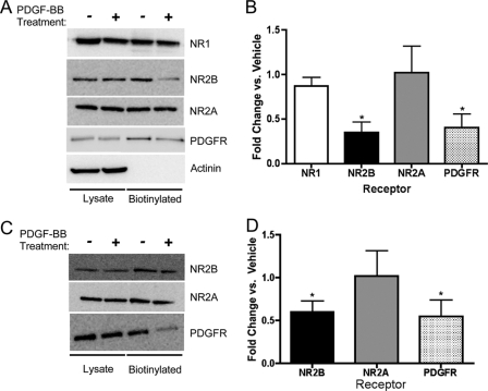
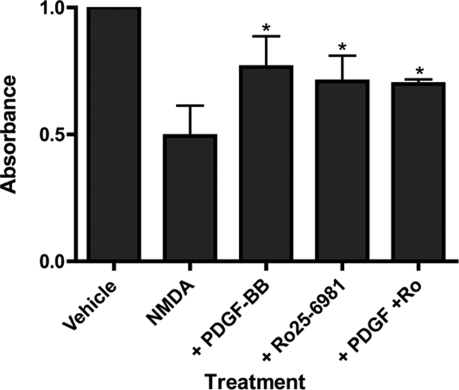
Platelet-derived growth factor (PDGF) beta receptor activation inhibits N-methyl-d-aspartate (NMDA)-evoked currents in hippocampal and cortical neurons via the activation of phospholipase Cgamma, PKC, the release of intracellular calcium, and a rearrangement of the actin cytoskeleton. In the hippocampus, the majority of NMDA receptors are heteromeric; most are composed of 2 NR1 subunits and 2 NR2A or 2 NR2B subunits. Using NR2B- and NR2A-specific antagonists, we demonstrate that PDGF-BB treatment preferentially inhibits NR2B-containing NMDA receptor currents in CA1 hippocampal neurons and enhances long-term depression in an NR2B subunit-dependent manner. Furthermore, treatment of hippocampal slices or cultures with PDGF-BB decreases the surface localization of NR2B but not of NR2A subunits. PDGFbeta receptors colocalize to a higher degree with NR2B subunits than with NR2A subunits. After neuronal injury, PDGFbeta receptors and PDGF-BB are up-regulated and PDGFbeta receptor activation is neuroprotective against glutamate-induced neuronal damage in cultured neurons. We demonstrate that the neuroprotective effects of PDGF-BB are occluded by the NR2B antagonist, Ro25-6981, and that PDGF-BB promotes NMDA signaling to CREB and ERK1/2. We conclude that PDGFbetaR signaling, by preferentially targeting NR2B receptors, provides an important mechanism for neuroprotection by growth factors in the central nervous system.


Figures

FIGURE 1.
FIGURE 1.
A, vehicle (0.0002% bovine serum albumin, 8 μm HCl) or 10 ng/ml PDGF-BB was applied to isolated CA1 hippocampal neurons (as indicated by the black bar). NMDA-evoked currents were recorded every 60 s by application of 1 μm NMDA, 0.5 μm glycine for 3 s. Sample traces from the same cell are shown with vehicle application (black trace) or with 10 ng/ml PDGF-BB application (gray trace). Data are representative of six experiments. B, NMDA-evoked currents were recorded every 60 s by application of 1 μm NMDA, 0.5 μm glycine for 3 s. Vehicle or PDGF-BB was applied to isolated CA1 hippocampal neurons in the presence of 500 nm Ro25-6981 (NR2B-selective antagonist). Sample traces from the same cell are shown with vehicle application (black trace) or with 10 ng/ml PDGF-BB application (gray trace). Data are representative of six experiments. C, NMDA-evoked currents were recorded every 60 s by application of 1 μm NMDA, 0.5 μm glycine for 3 s. Vehicle or PDGF-BB was applied to isolated CA1 hippocampal neurons in the presence of 50 nm NVP-AAM077 (NR2A-selective antagonist). Sample traces from the same cell are shown with vehicle application (black trace) or with 10 ng/ml PDGF-BB application (gray trace). Data are representative of seven experiments.
FIGURE 2.
FIGURE 2.
PDGF-BB application results in the selective reduction of the surface expression of NR2B. CA1 hippocampal slices were treated with vehicle or 10 μg/ml PDGF-BB for 10 min. The slices were biotinylated and lysates were incubated overnight with streptavidin beads. A, totallysates or biotinylated samples were blotted using antibodies against NR1 (n = 5), NR2B (n = 7), NR2A (n = 7, C), PDGFβ receptor (n = 4), or α-actinin (n = 4). B, PDGF-BB treatment decreases the surface localization of NR2B and PDGFβ receptors, but not NR1 or NR2A (*, p < 0.05 versus vehicle, Student's unpaired t tests). C and D, PDGF-BB treatment also decreased NR2B (n = 4) and PDGFβ receptor (n = 4) surface localization in hippocampal cultures (*, p < 0.05 versus vehicle, Student's unpaired t tests).
FIGURE 3.
FIGURE 3.
Acutely dissected hippocampal slices were incubated for 10 min with vehicle (control, -) or PDGF-BB 10 ng/ml (+). Lysates were resolved on SDS gels, transferred to nitrocellulose membranes, and probed with antibodies directed against phosphorylated tyrosine 1472. Membranes were stripped and reprobed for total NR2B (n = 4).
FIGURE 4.
FIGURE 4.
PDGFβ receptors are primarily extrasynaptic and colocalize with NR2B. A, merged image of PDGFβ receptor staining (green) and PSD-95 staining (red), including a high magnification image of one of the processes. Boxes represent regions of interest that were analyzed for Manders' overlap coefficient. The color scatter plot shows green, red, and yellow (overlap) pixels. The average Manders' overlap coefficient was 0.400. B, merged image of PDGFβ receptor staining (red) and NR2B staining (green), including a high magnification image of one of the processes. Boxes represent regions of interest that were analyzed for Manders' overlap coefficient. The color scatter plot shows green, red, and yellow (overlap) pixels. The average Manders' overlap coefficient was 0.808. C, merged image of PDGFβ receptor staining (red) and NR2A staining (green), including a high magnification image of one of the processes. Boxes represent regions of interest that were analyzed for Manders' overlap coefficient. The color scatter plot shows green, red, and yellow (overlap) pixels. The average Manders' overlap coefficient was 0.438. D, PDGFβ receptors show significantly higher colocalization with NR2B compared with NR2A or PSD-95. *, p < 0.05. Comparison of total pixel count between PDGFβ receptor images and between NR2A and NR2B were not significantly different (data not shown). E and F, comparison of total pixel count between PDGFβ receptor images and between NR2A and NR2B, non-parametric statistical comparison. PDGFβ receptor pixel numbers are not significantly different (p > 0.05) and NR2A and NR2B pixel numbers are not significantly different (p > 0.05).
FIGURE 5.
FIGURE 5.
PDGF-BB enhanced long-term depression in CA1 region of hippocampal slice. A, vehicle (0.0002% bovine serum albumin, 8 μm HCl) or PDGF-BB (10 ng/ml) was applied for 10 min after stable baseline recording as indicated by the open bar. Long-term depression was evoked by low-frequency stimulation after the vehicle treatment but the magnitude of LTD was increased after PDGF-BB treatment. B, PDGF-BB (10 ng/ml) or PDGF plus Ro25-6981 (0.5 μm) was applied for 10 min after stable baseline recording as indicated by the open bar. Ro25-6981 treatment did not block LTD, but PDGF-BB failed to facilitate LTD in the presence of Ro25-6981. Insets, representative traces recorded at the indicated time (numbers). Calibration bars, vertical = 0.25 mV; horizontal = 5 ms.
FIGURE 6.
FIGURE 6.
PDGF-BB is neuroprotective. Day 18-21 cultured hippocampal neurons were pretreated for 10 min with vehicle (ECF), 10 ng/ml PDGF-BB, 2.5 μm Ro25-6981, or both. Cells were subsequently incubated with vehicle (ECF) or 100 μm NMDA, 1 μm glycine for 3 min. ECF was aspirated and cells were returned to media for 24 h before determining cell viability as determined by enzyme-linked immunosorbent assay. Data represent the mean ± S.E. of four experiments (*, p < 0.05, analysis of variance with Bonferroni's post-test compared with NMDA-treated cells alone, second bar).
FIGURE 7.
FIGURE 7.
PDGF-BB treatment alters NMDA signaling to transcription factors in hippocampal neurons. For “total” NMDA treatment (A-C), 14-21-day-old hippocampal cultures were treated for 10 min in warm ECF with vehicle (-) or PDGF-BB (+), followed by treatment with vehicle or 100 μm NMDA and 1 μm glycine. All treatments were in the presence of 1 μm tetrodotoxin, 40 μm 6-cyano-7-nitroquinoxaline-2,3-dione, and 5 μm nifedipine. For “extrasynaptic” NMDA treatment (D-F), cultures were treated for 10 min with vehicle or PDGF-BB. During the last 3 min of the PDGF-BB treatment, 50 μm MK-801, 10 μm bicuculline, 10 μm glycine, and 5 μm nifedipine were included to block synaptic NMDA receptors. After either treatment, cells were washed briefly and incubated with vehicle or 100 μm NMDA, 10 μm glycine for 3 min in the presence of 6-cyano-7-nitroquinoxaline-2,3-dione and nifedipine. A, after total NMDA treatment, membranes were probed with anti-phospho-ERK, phospho-CREB, and ERK. Blots are representative of five to seven experiments and immunoreactivity was quantified versus control (vehicle, -)(B, C). D, after extrasynaptic NMDA treatment, membranes were probed with anti-phospho-ERK, ERK, and phospho-CREB. Blots are representative of five to seven experiments and immunoreactivity was quantified versus control (vehicle, -)(E, F). Phospho-ERK immunoreactivity was normalized to total ERK (both bands were pooled) and phospho-CREB immunoreactivity was normalized to total CREB (not shown).
FIGURE 8.
FIGURE 8.
The proposed mechanisms of PDGFβR inhibition of NMDA receptors and neuroprotection. PDGFβ receptor activation inhibits NR2B-containing NMDA receptors and selectively removes NR2B subunits from the cell membrane. However, the relationship between these two observations remains unclear. The removal of NR2B may cause, or be the result of, PDGFβ receptor inhibition of NMDA currents. PDGFβ receptor activation is also neuroprotective. PDGF-BB treatment led to an increase in ERK1/2 phosphorylation and prevented extrasynaptic NMDA receptor inhibition of ERK1/2 and CREB phosphorylation.

References

    1. Paoletti, P., and Neyton, J. (2007) Curr. Opin. Pharmacol. 7 39-47 - PubMed
    1. Groc, L., Heine, M., Cousins, S. L., Stephenson, F. A., Lounis, B., Cognet, L., and Choquet, D. (2006) Proc. Natl. Acad. Sci. U. S. A. 103 18769-18774 - PMC - PubMed
    1. Al-Hallaq, R. A., Conrads, T. P., Veenstra, T. D., and Wenthold, R. J. (2007) J. Neurosci. 27 8334-8343 - PMC - PubMed
    1. Tovar, K. R., and Westbrook, G. L. (2002) Neuron 34 255-264 - PubMed
    1. Groc, L., Choquet, D., Stephenson, F. A., Verrier, D., Manzoni, O. J., and Chavis, P. (2007) J. Neurosci. 27 10165-10175 - PMC - PubMed

Publication types

MeSH terms