. 2009 Jan 13;106(2):468-73.
doi: 10.1073/pnas.0809824105. Epub 2008 Dec 23.

Mice lacking asparaginyl endopeptidase develop disorders resembling hemophagocytic syndrome

Affiliations

Mice lacking asparaginyl endopeptidase develop disorders resembling hemophagocytic syndrome

Chi-Bun Chan et al. Proc Natl Acad Sci U S A. .

Erratum in

  • Proc Natl Acad Sci U S A. 2009 Apr 7;106(14):6023. Zheng, Chengyun [added]; Henter, Jan-Inge [added]; Meeths, Marie [added]; Nordenskjold, Magnus [added]

Abstract

Asparaginyl endopeptidase (AEP or legumain) is a lysosomal cysteine protease that cleaves protein substrates on the C-terminal side of asparagine. AEP plays a pivotal role in the endosome/lysosomal degradation system and is implicated in antigen processing. The processing of the lysosomal proteases cathepsins in kidney is completely defective in AEP-deficient mice with accumulation of macromolecules in the lysosomes, which is typically seen in lysosomal disorders. Here we show that mutant mice lacking AEP develop fever, cytopenia, hepatosplenomegaly, and hemophagocytosis, which are primary pathological manifestations of hemophagocytic syndrome/hemophagocytic lymphohistiocytosis (HLH). Moreover, AEP deficiency provokes extramedullary hematopoiesis in the spleen and abnormally enlarged histiocytes with ingested red blood cells (RBCs) in bone marrow. Interestingly, RBCs from AEP-null mice are defective in plasma membrane components. Further, AEP-null mice display lower natural killer cell activity, but none of the major cytokines is substantially abnormal. These results indicate that AEP might be a previously unrecognized component in HLH pathophysiology.


Conflict of interest statement

The authors declare no conflict of interest.

Figures
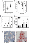
Fig. 1.
Fig. 1.
Higher body temperature and anemia in AEP-deficient mice. (A) Body temperature of AEP-knockout (filled circles) and control heterozygous (open circles) mice at 12 months of age. ***, P < 0.005, Student's t test, +/−, n = 4, −/−, n = 7. Body core temperature was continuously recorded on a potentiometer by a copper–constantan thermocouple covered with polyethylene tubing (outside diameter 1 mm). (B) Age-dependent decrease of circulating hematocrit was seen in AEP-null (filled circles) animals but not in the heterozygous control mice (open circles). *, P < 0.05, Student's t test, 3-month-old: +/−, n = 6, −/−, n = 6; 6-month-old: +/−, n = 7, −/−, n = 6; 15-month-old: +/−, n = 2, −/−, n = 4. (C) Age-dependent increase of circulating reticulocytes was seen in AEP-null (filled circles) animals but not in the heterozygous control mice (open circles) *, P < 0.05; **, P < 0.01, Student's t test). Data were obtained from the same mice examined in B. (D) Circulating Epo from wild-type (filled bars) and AEP-knockout (open bars) were determined by ELISA. A progressive increase of serum Epo was recorded in AEP-null animals with increased age. *, P < 0.05, Student's t test, n = 6. (E) Bone marrow sections from femur of wild-type (+/+) and AEP-knockout (−/−) animals were stained with EpoR-specific antibody. IHC, immunohistochemistry. Enhanced EpoR expression was detected in AEP-knockout mice. (Scale bar, 20 μm.)
Fig. 2.
Fig. 2.
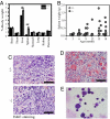
Hepatosplenomegaly in AEP-knockout mice. (A) The weights of both the liver and spleen from AEP-null animals (open bar) were significantly higher than those of age-matched wild-type controls (filled bar). *, P < 0.05; ***, P < 0.001, n = 8. (B) Age-dependent increase of spleen weight in AEP-knockout mice. AEP-knockout mice (filled circles) and heterozygous control (open circles) of various ages as indicated were killed and the spleens were weighed (3-month-old: +/−, n = 6, −/−, n = 6; 6-month-old: +/−, n = 6, −/−, n = 6; 9-month-old: +/−, n = 3, −/−, n = 10; 12-month-old: +/−, n = 4, −/−, n = 3; 15-month-old: +/−, n = 3, −/−, n = 10; 18-month-old: +/−, n = 6, −/−, n = 8.). (C) H&E staining of the spleen from both AEP-heterozygous (+/−) and homozygous mice (−/−). (Scale bar, 500 μm.) (D) H&E staining of AEP-null spleen showing numerous hemophagocytes. (Scale bar, 500 μm.) (E) Giemsa staining of splenocytes from AEP−/− mice showing immature myeloid lineage cells.
Fig. 3.
Fig. 3.
Extramedullary hematopoiesis of myeloid lineage in AEP-knockout mice. (A) Non-T and non-B cells increased in splenocytes with age and spleen weight in AEP-knockout mice. Spleen weight is shown above each graph. (B and C) A substantial increase of erythroid cells (Ter-119/CD45) (B) and small increase of myeloid cells (Gr-1/Mac-1) (C) occurred in spleen from AEP-null mice compared with wild-type mice.
Fig. 4.
Fig. 4.
Enhanced phagocytic activity against RBCs in AEP-null macrophages. (A) H&E staining of the bone marrow and spleen from wild-type and AEP-lacking mice. AEP-null mice display “Gaucher-like cells” in bone marrow with engulfed RBCs. The bone marrow and spleen from AEP-null mice contained numerous Gaucher-like cells. The histiocytes in AEP-lacking mice contained numerous engulfed RBCs (arrows). (Scale bar, 10 μm.) (B) Electron microscopic analysis of histiocytes in bone marrow from AEP-null animals. R, RBC. (Scale bar, 2 μm.) (C) Enhanced phagocytic activity of peritoneal macrophages from AEP-knockout mice. Peritoneal macrophages from 2-month-old AEP-null mice (−/−) and control heterozygous mice (+/−) were used for the phagocytosis assay with fluorescent microspheres. (Left) Representative results. (Right) Results are shown as a histogram; data are expressed as mean ± SD. *, P < 0.05, Student's t test, n = 6. (D) Peritoneal macrophages from AEP-knockout mice (−/−) and control heterozygous mice (+/−) were used for the phagocytosis assay against RBCs from AEP-knockout mice and control heterozygous mice (+/−). RBCs were untreated (−) or opsonized (+) with anti-mouse RBC antibody before the assay. Macrophages (Mφ) and RBCs were taken from 12-month-old mice and incubated for 1 h. Data are expressed as mean ± SD. ***, P < 0.001 against untreated group; **, P < 0.001 against opsonized group in different genotype using the same RBCs, Student's t test, n = 6. (E) Reduced natural killer cells activity in AEP-knockout mice. Percent cytotoxicity of NK cells against P815 mastocytoma from aged-matched wild-type (filled bar) and AEP-knockout (open bar) were measured by flow cytometry. Results are expressed as mean ± SEM. *, P < 0.05, Student's t test, n = 3.

References

    1. Janka GE. Familial and acquired hemophagocytic lymphohistiocytosis. Eur J Pediatr. 2007;166:95–109. - PubMed
    1. Janka GE. Hemophagocytic syndromes. Blood Rev. 2007;21:245–253. - PubMed
    1. Stepp SE, et al. Perforin gene defects in familial hemophagocytic lymphohistiocytosis. Science. 1999;286:1957–1959. - PubMed
    1. Feldmann J, et al. Munc13-4 is essential for cytolytic granules fusion and is mutated in a form of familial hemophagocytic lymphohistiocytosis (FHL3) Cell. 2003;115:461–473. - PubMed
    1. Menasche G, et al. Mutations in RAB27A cause Griscelli syndrome associated with haemophagocytic syndrome. Nat Genet. 2000;25:173–176. - PubMed

Publication types

Substances

LinkOut - more resources