. 2008 Dec;20(12):3273-88.
doi: 10.1105/tpc.107.057489. Epub 2008 Dec 23.

A tobacco calcium-dependent protein kinase, CDPK1, regulates the transcription factor REPRESSION OF SHOOT GROWTH in response to gibberellins

Affiliations

A tobacco calcium-dependent protein kinase, CDPK1, regulates the transcription factor REPRESSION OF SHOOT GROWTH in response to gibberellins

Sarahmi Ishida et al. Plant Cell. 2008 Dec.

Abstract

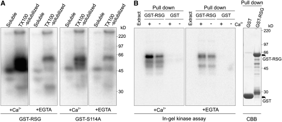
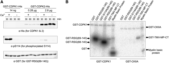
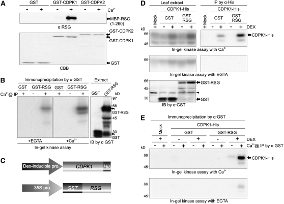
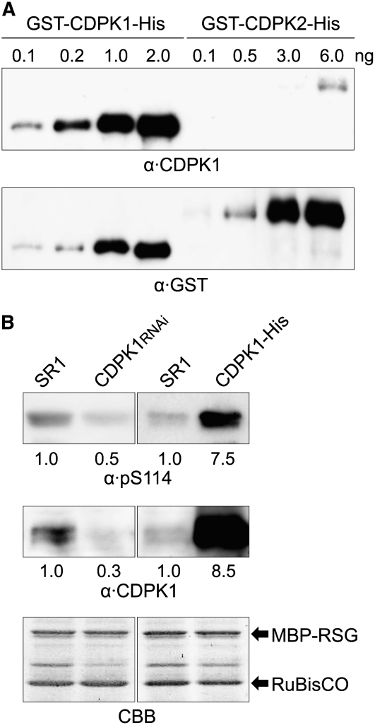
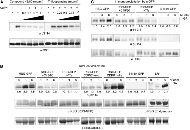
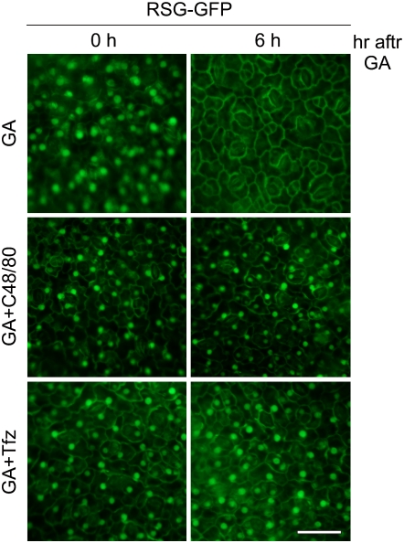
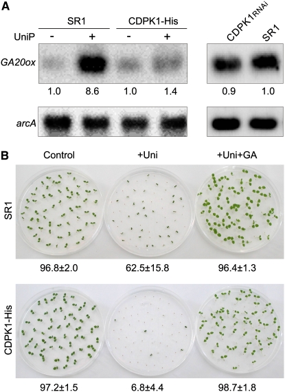
The homeostasis of gibberellins (GAs) is maintained by negative feedback in plants. REPRESSION OF SHOOT GROWTH (RSG) is a tobacco (Nicotiana tabacum) transcriptional activator that has been suggested to play a role in GA feedback by the regulation of GA biosynthetic enzymes. The 14-3-3 signaling proteins negatively regulate RSG by sequestering it in the cytoplasm in response to GAs. The phosphorylation on Ser-114 of RSG is essential for 14-3-3 binding of RSG. Here, we identified tobacco Ca(2+)-dependent protein kinase (CDPK1) as an RSG kinase that promotes 14-3-3 binding to RSG by phosphorylation of Ser-114 of RSG. CDPK1 interacts with RSG in a Ca(2+)-dependent manner in vivo and in vitro and specifically phosphorylates Ser-114 of RSG. Inhibition of CDPK repressed the GA-induced phosphorylation of Ser-114 of RSG and the GA-induced nuclear export of RSG. Overexpression of CDPK1 inhibited the feedback regulation of a GA 20-oxidase gene and resulted in sensitization to the GA biosynthetic inhibitor. Our results suggest that CDPK1 decodes the Ca(2+) signal produced by GAs and regulates the intracellular localization of RSG.


Figures

Figure 1.
Figure 1.
RSG Kinase Activities in Tobacco. (A) Enhancement of RSG kinase activities in the detergent-solubilized fraction by Ca2+. Tobacco leaf cell extract was fractioned into a detergent-free soluble fraction (Soluble) and a Triton X-100–solubilized fraction (TX100-solubilized) followed by an in-gel kinase assay with recombinant RSG (GST–RSG) or its S114A mutant version (GST–S114A) as substrates in the presence or absence of Ca2+ (+Ca2+ or +EGTA). The same proportion of the detergent-free soluble and the Triton X-100–solubilized fractions of the plant proteins were loaded on the gel. (B) Enhancement of the interaction between RSG kinase and RSG by Ca2+. Tobacco leaf cell extract was incubated with glutathione beads immobilized with GST or GST–RSG in the presence (+) or the absence (−) of Ca2+ as indicated. The precipitates with beads (Pull down) and the leaf cell extract (Extract) were subjected to an in-gel kinase assay with acrylamide gels containing the GST-tagged phosphorylation domain of RSG [residues 69 to 140, GST–RSG(69-140)] as a substrate with or without Ca2+ (+Ca2+ or +EGTA). The precipitates were also subjected to SDS-PAGE (Tris/Glycine buffer) and stained by Coomassie blue (CBB; right panel). The experiments were repeated twice with similar results.
Figure 2.
Figure 2.
Phosphorylation of RSG by CDPK1 in Vitro. (A) Comparison of the catalytic activities of CDPK1 and CDPK2 on RSG. The GST-tagged phosphorylation domain of RSG [residues 69-140, GST–RSG(69-140)] was phosphorylated by different concentrations of recombinant CDPK1 and CDPK2 (GST–CDPK1–His and GST–CDPK2–His, respectively) for indicated periods of time with or without Ca2+. Aliquots of reactions were subjected to SDS-PAGE (Tris/Glycine buffer) followed by immunoblot analysis with anti-pentad histidine (α·His, to detect CDPKs, top), anti-pS114, which specifically recognizes phosphorylated Ser-114 of RSG (α·pS114, middle), and anti-GST [α·GST, to detect GST–RSG(69-140), bottom]. Note that the recombinant CDPK1 amounts were too low for simultaneous detection with recombinant CDPK2. The enzymatic activity of recombinant CDPKs were confirmed using an in vitro phosphorylation assay on the general substrates myelin basic protein or casein. (B) Comparison of the catalytic activities of CDPK1 and Arabidopsis CKIIA on RSG. Control GST, GST-tagged phosphorylation domains of RSG [GST–RSG(69-140), GST–S114A(69-140), and GST–RSG(88-140)], GST-tagged tobacco mosaic virus–derived polypeptide (GST–TMV-MP-CT), and myelin basic protein were phosphorylated by recombinant Nt CDPK1 or recombinant At CKIIA (GST–CDPK1 and GST–CKIIA, respectively) in the presence of [γ-32P]ATP. Equal amounts of substrates were subjected to SDS-PAGE (Tris/Glycine buffer), and 32P-labeled proteins were visualized with a phosphor imager. The experiments were repeated twice with similar results.
Figure 3.
Figure 3.
Interaction between CDPK1 and RSG in Vitro and in Vivo. (A) RSG interacts with CDPK1 in a Ca2+-dependent manner but not with CDPK2 in vitro. Control GST and GST-tagged recombinant CDPKs (GST–CDPK1 or GST–CDPK2) were immobilized to glutathione beads and incubated with MBP-tagged recombinant RSG [residues 1 to 260, MBP–RSG(1-260)] in the presence (+) or the absence (−) of Ca2+ as indicated. After the binding reactions, glutathione bead-bound proteins were subjected to SDS-PAGE (Tris/Glycine buffer) followed by immunoblot analysis with anti-RSG (top) or Coomassie blue (CBB) staining (bottom). (B) RSG interacts with CDPKs in plant cells. Agrobacterium carrying the expression construct for GST–RSG or control GST driven by the 35S promoter of CaMV was infiltrated into tobacco leaves. Two days after infiltration, leaf extracts were prepared. The expression of GST fusion proteins was determined by immunoblot with the anti-GST antibody (IB by α·GST). The RSG-interacting proteins were immunoprecipitated by anti-GST in the presence (+) or absence (−) of Ca2+ as indicated (Ca2+ @ IP) and subjected to an in-gel kinase assay with the GST-tagged phosphorylation domain of RSG [residues 69 to 140, GST–RSG(69-140)] as a substrate in the presence or absence of Ca2+ (+Ca2+ or +EGTA box of In-gel kinase assay, respectively). (C) Constructs of DEX-inducible CDPK1–His and 35S-driven GST–RSG. pro., promoter. (D) Coexpression of CDPK1–His and GST–RSG by agroinfiltration. The expression constructs shown in (C) were introduced simultaneously into tobacco leaves by Agrobacterium-mediated infiltration. One day after infiltration, double-infiltrated leaves and control plants (Mock) were sprayed with DEX or water as indicated, and leaf cell extracts were prepared 1 d after the treatment. The leaf cell extract and immunoprecipitates by anti-pentad histidine in the presence of Ca2+ (IP by α·His) were subjected to an in-gel kinase assay with GST–RSG(69-140) as a substrate with or without Ca2+ (In-gel kinase assay with Ca2+ or EGTA, respectively). The minor 50-kD band might be caused by proteolytic degradation and/or posttranslational modifications. The expression of GST and GST–RSG was detected by immunoblot analysis with the anti-GST antibody (IB by α·GST). The arrows indicate an unknown tobacco endogenous protein that interacted with anti-GST. (E) RSG interacts with CDPK1 in plant cells. The double-infiltrated plants or control plants (Mock) were treated with DEX or water, and leaf cell extracts were prepared as described in (D). RSG-interacting proteins were immunoprecipitated from the leaf cell extracts with anti-GST in the presence or absence of Ca2+ (Ca2+ @ IP by α·GST) and subjected to an in-gel kinase assay with GST–RSG(69-140) as a substrate with or without Ca2+ (In-gel kinase assay with Ca2+ or EGTA, respectively). The experiments were repeated twice with similar results.
Figure 4.
Figure 4.
CDPK1 Is a Major Kinase for Ser-114 of RSG. (A) Anti-CDPK1 specifically recognizes CDPK1. Recombinant CDPK1 and CDPK2 (GST–CDPK1–His and GST–CDPK2–His, respectively) were blotted onto membrane as indicated and immunodetected with anti-CDPK1 and anti-GST. (B) The leaf cell extracts from control wild-type SR1, CDPK1 RNAi transgenic plants, or CDPK1–His–overexpressing transgenic plants were incubated with MBP-tagged recombinant RSG (MBP–RSG) in an in vitro phosphorylation buffer. Aliquots of the reactions were subjected to SDS-PAGE (Tris/Glycine buffer) followed by immunoblot analysis with anti-pS114 (top) or anti-CDPK1 (middle). Images of Coomassie blue (CBB) staining were used as a loading control (bottom). The values at the bottoms of the panels indicate the relative level of strengths of signals after standardization. The values of SR1 were set to 1.0. The experiments were repeated twice with similar results. SR1, control wild-type SR1 tobacco plants; CDPK1rnai, transgenic plants in which expression of CDPK1 was decreased by RNAi; CDPK1–His, transgenic plants overexpressing CDPK1–His under the control of the CaMV 35S promoter.
Figure 5.
Figure 5.
The Calmodulin Antagonists Suppress GA-Induced Phosphorylation of Ser-114 and 14-3-3 Binding to RSG. (A) The calmodulin antagonist Compound 48/80 and Trifluoperazine inhibit the catalytic activity of CDPK1 in vitro. The recombinant RSG [GST–RSG(69-140)] was phosphorylated by recombinant CDPK1 (GST–CDPK1–His) in vitro in the presence of Compound 48/80 or Trifluoperazine at the concentrations as indicated. Reactions were subjected to immunoblot analysis with anti-pS114 (top) or anti-GST (bottom). (B) Compound 48/80 and Trifluoperazine inhibit the GA-induced phosphorylation of Ser-114 of RSG. Tobacco plants were treated with the GA biosynthesis inhibitor Uniconazole P for 1 week to reduce endogenous GA and then sprayed with a GA3 solution or a GA3 solution containing the calmodulin antagonist Compound 48/80 or Trifluoperazine. At the time points indicated after GA3 application, leaves were harvested, and total leaf cell extracts were prepared. Aliquots of leaf cell extract were separated by SDS-PAGE (Tris/Glycine buffer) followed by immunoblot analysis with anti-pS114 (top) or anti-RSG (middle). Both anti-RSG antibodies and pS114 antibodies recognized multiple bands of GFP fusion proteins in the immunoblotting. Although the exact nature of the proteins is unknown, multiple bands could be caused by proteolytic degradation and/or posttranslational modifications. Images of Coomassie blue (CBB) staining (ribulose-1,5-bisphosphate carboxylase/oxygenase [Rubisco]) were demonstrated to indicate RSG is not degraded by GA3 treatment (bottom). The values at the bottom of the top panels indicate the relative level of strengths of signals after standardization using signals of RSG–GFP (transgenic panels) or RSG (SR1 panel) as a control. The values of signals at 0 h were set to 1.0. The experiments were repeated five times with similar results. (C) Compound 48/80 and Trifluoperazine suppress GA-induced interaction between RSG and 14-3-3. RSG-interacting proteins were immunoprecipitated with anti-GFP from the leaf cell extract prepared as in (B) and separated by SDS-PAGE (Tris/Glycine buffer) followed by immunoblot analysis with anti-14-3-3 (top), anti-pS114 (middle), or anti-RSG (bottom). The values at the bottoms of the anti-14-3-3 panels indicate the relative level of strengths of signals after standardization using signals of immunoprecipitated RSG–GFP as a loading control. The values of signals at 0 h were set to 1.0. The experiments were repeated five times with similar results. RSG–GFP, GA-treated transgenic plants expressing RSG–GFP under the control of the CaMV 35S promoter; RSG–GFP + C48/80, GA- and Compound 48/80–treated transgenic plants expressing RSG–GFP under the control of the CaMV 35S promoter; RSG–GFP + Tfz, GA- and Trifluoperazine-treated transgenic plants expressing RSG–GFP under the control of the CaMV 35S promoter; S114A–GFP, GA-treated transgenic plants expressing S114A–GFP under the control of the CaMV 35S promoter; RSG–GFP CDPK1rnai, GA-treated RSG–GFP transgenic plants in which expression of CDPK1 was decreased by RNAi; RSG–GFP CDPK1–His, GA-treated transgenic plants expressing RSG–GFP and CDPK1–His under the control of the CaMV 35S promoter; SR1, GA-treated control wild-type SR1 plants.
Figure 6.
Figure 6.
The Calmodulin Antagonists Repress GA-Induced Nuclear Export of RSG. Transgenic tobacco expressing RSG–GFP was treated with the GA biosynthesis inhibitor Uniconazole P for 1 week to reduce endogenous GA and then sprayed with a GA3 solution or a GA3 solution containing the calmodulin antagonist Compound 48/80 or Trifluoperazine. Leaves were harvested at the time points indicated, and fluorescence due to GFP was observed with an epifluorescence microscope. GA, GA-treated transgenic plants expressing RSG–GFP under the control of the CaMV 35S promoter; GA + C48/80, GA- and Compound 48/80–treated transgenic plants expressing RSG–GFP under the control of the CaMV 35S promoter; GA + Tfz, GA- and Trifluoperazine-treated transgenic plants expressing RSG–GFP under the control of the CaMV 35S promoter. Bar = 50 μm.
Figure 7.
Figure 7.
CDPK1 Was Modified as a Consequence of the GA Signal. (A) Protein gel blot analysis of the endogenous CDPK1. SR1 plants were treated with Uniconazole P for 1 week and then sprayed with a GA3 solution. At the indicated time points after GA3 application, leaves were harvested, and total leaf cell extracts were prepared. The levels of CDPK1 protein in each total leaf cell extract were visualized by immunoblot with anti-CDPK1 (top). Images of Coomassie blue (CBB) staining (Rubisco) were used as a loading control (bottom). The values at the bottom of the top panel indicate the relative level of strengths of signals after standardization using CBB staining of Rubisco as a loading control. The values of signals at 0 h were set to 1.0. The experiments were repeated twice with similar results. (B) The intracellular localization pattern of CDPK1 was not apparently affected by GA treatment. Top panel: Transgenic plants expressing CDPK1–His under the control of the CaMV 35S promoter were treated as in (A), and cells were fractionated to the Triton X-100–solubilized fraction (TX100-solubilized) and the detergent-free soluble fraction (Soluble). The same proportion of the detergent-free soluble and the Triton X-100–solubilized fractions of the plant proteins were loaded on the gel. The levels of CDPK1 were visualized by immunoblots with anti-CDPK1. Each value at the bottom of the panel indicates the relative level of strength of signal in a soluble fraction when the value of signal in a Triton X-100–solubilized fraction was set to 1.0. The experiments were repeated three times with similar results. Bottom panels: Transgenic plants expressing CDPK1–GFP under the control of the CaMV 35S promoter were treated as in (A), and fluorescence due to GFP was observed with an epifluorescence microscope at the indicated time points after GA3 application. Bar = 50 μm. (C) GA induces the phosphorylation of CDPK1. Aliquots of the Triton X-100–solubilized fraction prepared as in (B) were treated with or without a phosphatase (CIAP and Control, respectively) at 30°C for 1 h. Reactions were separated by SDS-PAGE (Tris/Glycine buffer) and subjected to immunoblot analysis with anti-CDPK1. To facilitate the detection of mobility shift of CDPK1, electrophoresis was performed using a gel of low concentration of acrylamide with extended running time. CIAP, calf intestine alkali phosphatase. The experiments were repeated five times with similar results. (D) The GA signal promotes the interaction between CDPK1 and RSG. Transgenic plants expressing either CDPK1–His and RSG–GFP under the control of the CaMV 35S promoter or RSG–GFP only under the control of the CaMV 35S promoter were treated as in (A). At the indicated time points after GA3 application, leaves were harvested, and leaf cell extracts were prepared. RSG-interacting proteins were immunoprecipitated with anti-RSG and analyzed by immunoblots with anti-CDPK1 (top) and anti-RSG (bottom). The values at the bottom of the top panels indicate the relative level of strengths of signals after standardization using signals of immunoprecipitated RSG–GFP as a loading control. The values of signals at 0 h were set to 1.0. The experiments were repeated five times with similar results.
Figure 8.
Figure 8.
Overexpression of CDPK1 Inhibited GA Homeostasis. (A) Feedback regulation of the GA 20-oxidase gene was suppressed in transgenic plants overexpressing CDPK1–His. Transgenic tobacco overexpressing CDPK1–His under the control of the CaMV 35S promoter and control wild-type SR1 plants were grown with or without the GA biosynthesis inhibitor Uniconazole P for 1 week as indicated. Transgenic tobacco in which expression of CDPK1 was repressed by RNAi and control wild-type SR1 plants were grown without chemical treatment. The mRNA levels of GA 20-oxidase (GA20ox) were examined by quantitative RT-PCR. After PCR, the products were detected by DNA gel blot hybridization. Tobacco arcA was amplified in the same reaction and used as an internal control for RT-PCR. The values at the bottom of the top panel indicate the relative level of strengths of signals after standardization using signals of arcA as a loading control. The values of SR1 were set to 1.0. The experiments were repeated three times with similar results. (B) Uniconazole P resistance was reduced in transgenic plants overexpressing CDPK1. Seeds of transgenic tobacco overexpressing CDPK1–His and control wild-type SR1 plants were germinated in a medium containing 1 μM Uniconazole P with or without 1 μM GA3 for 7 d at 28°C. The values at the bottom of panels indicate the germination ratio calculated from three independent plates (±sd, 80 to 100 seeds/plate). The experiments were repeated three times with similar results. SR1, control wild-type SR1 tobacco plants; CDPK1–His, transgenic tobacco plants overexpressing CDPK1–His under the control of the CaMV 35S promoter; CDPK1rnai, transgenic plants in which expression of CDPK1 was decreased by RNAi; +UniP, seedlings germinated on agarose containing Uniconazole P; +Uni+GA, seedlings germinated on agarose containing Uniconazole P and GA3.

References

    1. Allen, G.J., Chu, S.P., Harrington, C.L., Schumacher, K., Hoffmann, T., Tang, Y.Y., Grill, E., and Schroeder, J.I. (2001). A defined range of guard cell calcium oscillation parameters encodes stomatal movements. Nature 411 1053–1057. - PubMed
    1. Amador, V., Monte, E., García-Martínez, J.-L., and Prat, S. (2001). Gibberellins signal nuclear import of PHOR1, a photoperiod-responsive protein with homology to Drosophila armadillo. Cell 106 343–354. - PubMed
    1. Aoyama, T., and Chua, N.-H. (1997). A glucocorticoid-mediated transcriptional induction system in transgenic plants. Plant J. 11 605–612. - PubMed
    1. Arabidopsis Genome Initiative (2000). Analysis of the genome sequence of the flowering plant Arabidopsis thaliana. Nature 408 796–815. - PubMed
    1. Asano, T., et al. (2002). Rice SPK, a calmodulin-like domain protein kinase, is required for storage product accumulation during seed development: phosphorylation of sucrose synthase is a possible factor. Plant Cell 14 619–628. - PMC - PubMed

Publication types

MeSH terms