

Review
doi: 10.1161/CIRCULATIONAHA.108.767012.
Takotsubo cardiomyopathy: a new form of acute, reversible heart failure
Affiliations
- PMID: 19106400
- PMCID: PMC4893309
- DOI: 10.1161/CIRCULATIONAHA.108.767012
Item in Clipboard
Review
Takotsubo cardiomyopathy: a new form of acute, reversible heart failure
Circulation.
.
No abstract available
Figures
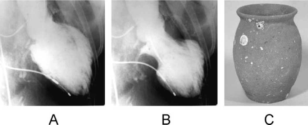
Left ventriculogram (A, end-diastolic phase; B, end-systolic phase) in the right anterior oblique projection. The extensive area around the apex shows akinesis, and the basal segments display hypercontraction, especially in the end-diastolic phase. C, A picture of a real takotsubo, which has a round bottom and narrow neck to capture octopuses and has been used for a long time in Japan.
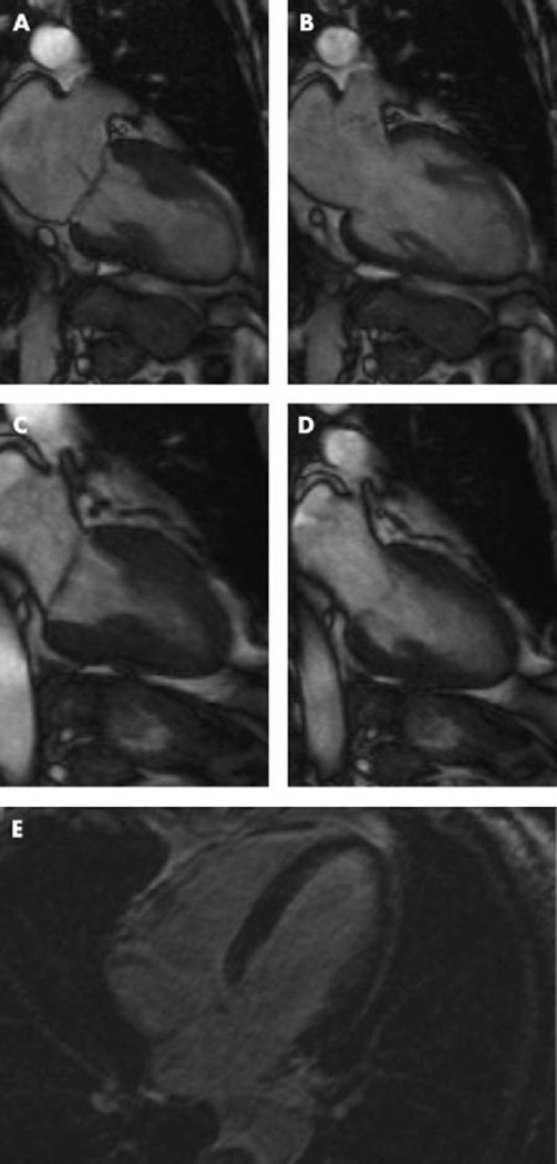
Cine sequences of cardiac magnetic resonance imaging during systole (A) and diastole (B) in the acute phase. Normal function could be documented after 3 weeks (C, systole; D, diastole). Contrast-enhanced cardiovascular magnetic resonance image did not show myocardial hyperenhancement even in the delayed phase (E). Adapted from Nef et al, with permission.
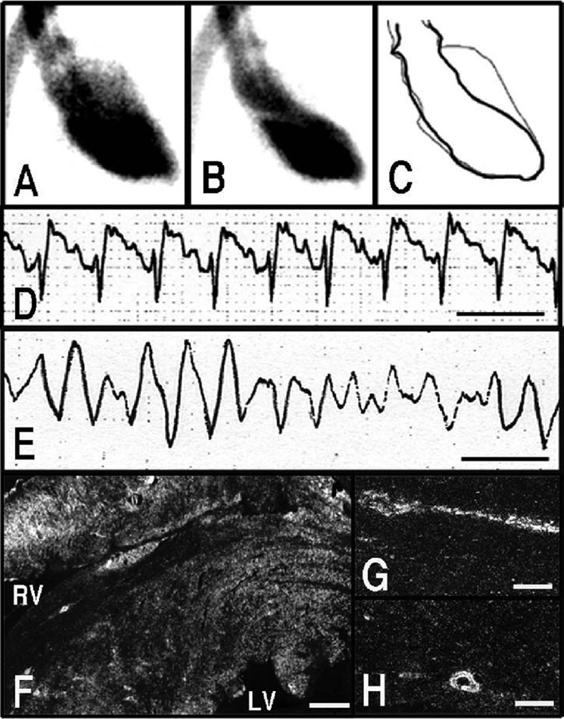
Left ventriculographic (A through C) and ECG (D and E) changes in response to immobilization of rats. Left ventriculogram, right anterior oblique (30°) projection. A, Diastole. B, systole. C, The trace of A and B. Reduced left ventricular contraction around the left ventricular apex was observed in response to stress. ECG, lead II. Line indicates 0.2 second. ST-segment elevation (D) was observed in response to stress. A case of ventricular fibrillation also was observed (E). Dark-field photomicrograph showing signals for c-jun mRNA in the heart (F) and coronary arteries (G and H) sampled at 30 minutes from the onset of immobilization. Strong signals were observed in myocardium surrounding the left and right ventricular cavities (F). These signals also were observed in endothelial cells and smooth muscle cells of coronary arteries (G and H). Bar=600 µm (F) and 100 µm (G and H). A through C, Adapted and modified from Ueyama et al, with permission from the Japanese Circulation Society.
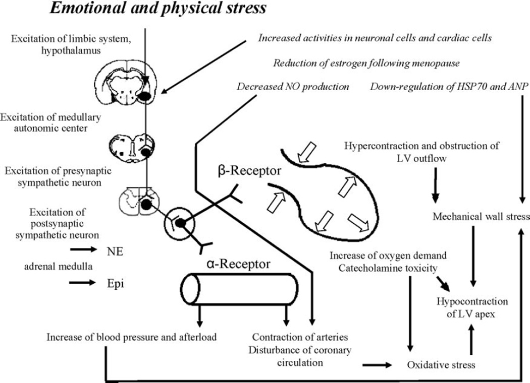
Possible underlying mechanism of classic takotsubo cardiomyopathy. See text for details.
References
-
- Tsuchihashi K, Ueshima K, Uchida T, Oh-Mura N, Kimura K, Owa M, Yoshiyama M, Miyazaki S, Haze K, Ogawa H, Honda T, Hase M, Kai R, Morii I. Transient left ventricular apical ballooning without coronary artery stenosis: a novel heart syndrome mimicking acute myocardial infarction: Angina Pectoris-Myocardial Infarction Investigations in Japan. J Am Coll Cardiol. 2001;38:11–18. - PubMed
-
- Kurisu S, Sato H, Kawagoe T, Ishihara M, Shimatani Y, Nishioka K, Kono Y, Umemura T, Nakamura S. Tako-tsubo-like left ventricular dysfunction with ST-segment elevation: a novel cardiac syndrome mimicking acute myocardial infarction. Am Heart J. 2002;143:448–455. - PubMed
-
- Akashi YJ, Musha H, Kida K, Itoh K, Inoue K, Kawasaki K, Hashimoto N, Miyake F. Reversible ventricular dysfunction takotsubo cardiomyopathy. Eur J Heart Fail. 2005;7:1171–1176. - PubMed
-
- Sato M, Fujita S, Saito A, Ikeda Y, Kitazawa H, Takahashi M, Ishiguro J, Okabe M, Nakamura Y, Nagai T, Watanabe H, Kodama M, Aizawa Y. Increased incidence of transient left ventricular apical ballooning (socalled “Takotsubo” cardiomyopathy) after the mid-Niigata Prefecture earthquake. Circ J. 2006;70:947–953. - PubMed
-
- Iga K, Gen H, Tomonaga G, Matsumura T, Hori K. Reversible left ventricular wall motion impairment caused by pheochromocytoma: a case report. Jpn Circ J. 1989;53:813–818. - PubMed
Publication types
MeSH terms
Grants and funding
LinkOut - more resources
Full Text Sources
Other Literature Sources
Medical
