. 2009 Feb 13;104(3):346-54.
doi: 10.1161/CIRCRESAHA.108.185785. Epub 2008 Dec 23.

Stimulation of Toll-like receptor 2 in human platelets induces a thromboinflammatory response through activation of phosphoinositide 3-kinase

Affiliations

Stimulation of Toll-like receptor 2 in human platelets induces a thromboinflammatory response through activation of phosphoinositide 3-kinase

Price Blair et al. Circ Res. .

Abstract

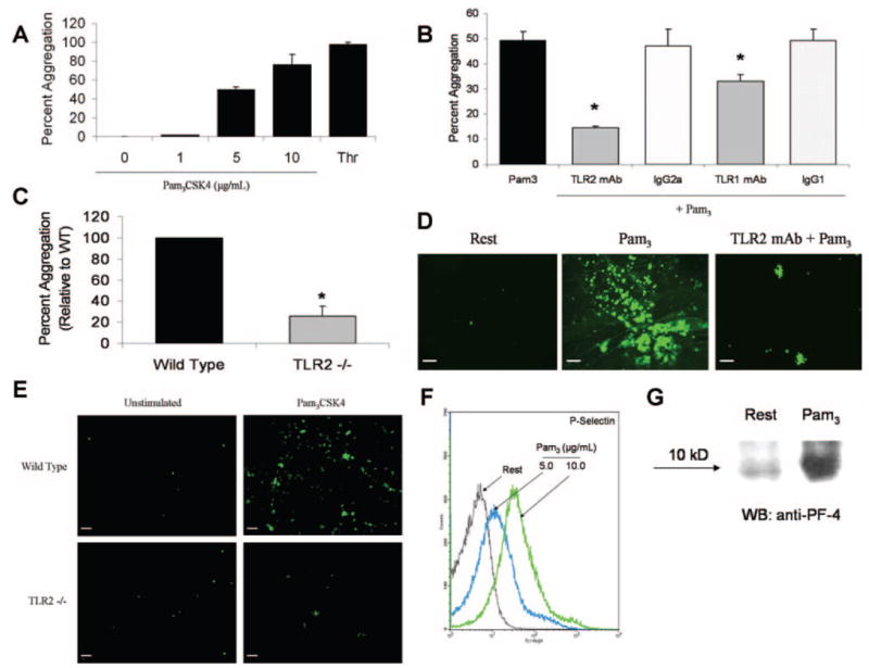
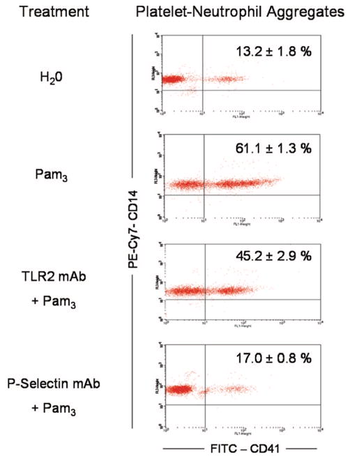
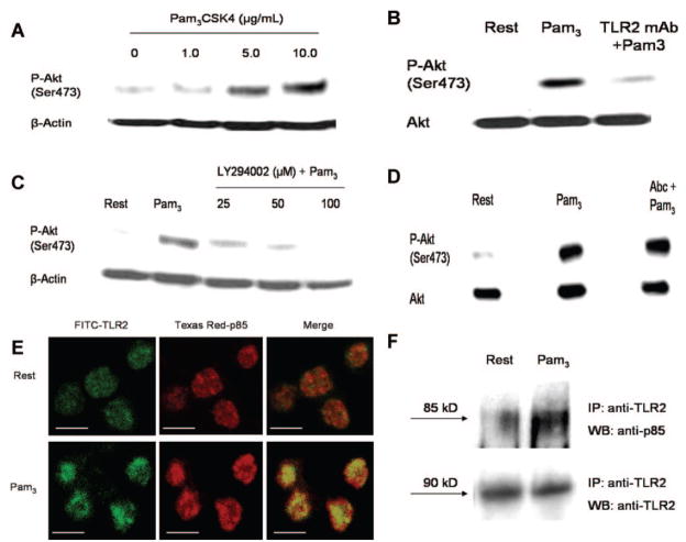
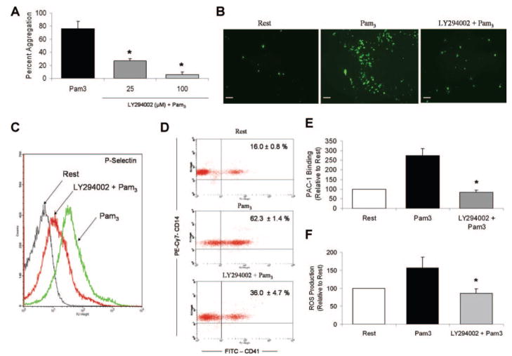
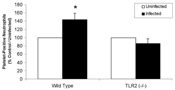
Cells of the innate immune system use Toll-like receptors (TLRs) to initiate the proinflammatory response to microbial infection. Recent studies have shown acute infections are associated with a transient increase in the risk of vascular thrombotic events. Although platelets play a central role in acute thrombosis and accumulating evidence demonstrates their role in inflammation and innate immunity, investigations into the expression and functionality of platelet TLRs have been limited. In the present study, we demonstrate that human platelets express TLR2, TLR1, and TLR6. Incubation of isolated platelets with Pam(3)CSK4, a synthetic TLR2/TLR1 agonist, directly induced platelet aggregation and adhesion to collagen. These functional responses were inhibited in TLR2-deficient mice and, in human platelets, by pretreatment with TLR2-blocking antibody. Stimulation of platelet TLR2 also increased P-selectin surface expression, activation of integrin alpha(IIb)beta(3), generation of reactive oxygen species, and, in human whole blood, formation of platelet-neutrophil heterotypic aggregates. TLR2 stimulation also activated the phosphoinositide 3-kinase (PI3-K)/Akt signaling pathway in platelets, and inhibition of PI3-K significantly reduced Pam(3)CSK4-induced platelet responses. In vivo challenge with live Porphyromonas gingivalis, a Gram-negative pathogenic bacterium that uses TLR2 for innate immune signaling, also induced significant formation of platelet-neutrophil aggregates in wild-type but not TLR2-deficient mice. Together, these data provide the first demonstration that human platelets express functional TLR2 capable of recognizing bacterial components and activating the platelet thrombotic and/or inflammatory pathways. This work substantiates the role of platelets in the immune and inflammatory response and suggests a mechanism by which bacteria could directly activate platelets.


Figures

Figure 1
Figure 1
Pam3CSK4 induces platelet aggregation, adhesion, and secretion in a TLR2-dependent manner. A and B, Pam3CSK4 (1 to 10 μg/mL) or thrombin (0.5 U/mL) were added to stirred human platelets, and aggregation was monitored for 6 minutes. Several platelet suspensions (B) were preincubated with functional grade anti-TLR2 mAb, anti-TLR1 mAb, or equal concentrations of their respective isotype controls (25 μg/mL) before stimulation with Pam3CSK4 (5 μg/mL). Data are reported as maximum percentage of aggregation±SD (n=3) for all groups. *P<0.01 compared to Pam3CSK4 alone. C, Pam3CSK4 (30 μg/mL) was added to stirred murine platelets isolated from either control C57BL/6 or TLR2-deficient mice. Aggregation was monitored for 6 minutes, and maximum percentage of aggregation was recorded. Reported data are normalized to control/wild-type (n=5 for both groups). *P<0.001 compared to control/wild-type. D, Calcein-labeled human platelets (resting or Pam3CSK4-stimulated [10 μg/mL]) were recirculated over collagen-covered coverslips for 20 minutes in a cell adhesion flow chamber. For some studies, platelets were pretreated with TLR2-blocking antibody (25 μg/mL) before stimulation. Adherent cells were viewed on a fluorescent microscope at X20 magnification. Representative fluorescent micrographs are shown. Scale bar=5 μm. E, Calcein-labeled platelets isolated from control C57BL/6 or TLR2-deficient mice were left quiescent or stimulated with Pam3CSK4 (10 μg/mL) and recirculated over collagen-coated coverslips for 20 minutes. Representative fluorescent micrographs are shown. Scale bar=5 μm. F, Washed human platelets (resting or Pam3CSK4-stimulated) were labeled with FITC–anti–P-selectin mAb and analyzed by flow cytometry. A representative histogram of 3 independent experiments is shown. G, Isolated human platelets were left quiescent or stimulated with Pam3CSK4 (10 μg/mL). The supernatant/platelet releasate was harvested, and equal volumes of releasate protein were separated by gel electrophoresis and immunoblotted for platelet factor-4 (PF-4).
Figure 2
Figure 2
Pam3CSK4 does not induce platelet aggregation in human whole blood but stimulates formation of platelet–leukocyte aggregates. A, Human whole blood was diluted in 0.9% saline and stirred in a whole blood lumi-aggregometer. Pam3CSK4 (10 μg/mL) was added, and aggregation was monitored for 10 minutes. Some samples were treated with TLR2-blocking antibody (25 μg/mL) or P-selectin–blocking antibody (10 μg/mL) before stimulation. Immediately following aggregation, 30 μL of whole blood was extracted and added to a cocktail containing FITC–anti-CD41 and PE-Cy7–anti-CD14 mAbs. Samples were analyzed by flow cytometry. Dot plots representative of 3 independent experiments are shown. Events located in the upper right quadrant represent CD14-positive/CD41-positive neutrophils. The percentage of collected, platelet-positive neutrophils was quantified (n=3 for all groups).
Figure 3
Figure 3
Pam3CSK4 stimulation of human platelets activates Akt in a TLR2/PI3-K-dependent manner. A through D, Platelets were left quiescent or stimulated with increasing concentrations of Pam3CSK4 (1 to 10 μg/mL) (A). Alternatively, platelets were treated with TLR2-blocking antibody (25 μg/mL) (B), LY294002 (25 to 100 μmol/L) (C), or abciximab (10 μg/mL) (D) before stimulation with Pam3CSK4 (10 μg/mL). For abciximab-treated samples, platelets were supplemented with fibrinogen (1 mg/mL) and stirred constantly. Equal amounts of platelet homogenate were resolved by SDS-PAGE and immunoblotted for phospho-Akt(Ser473), AKT, or β-actin. Representative blots are shown. E, Resting or Pam3CSK4-stimulated (10 μg/mL) platelets were stained with anti-TLR2 (green) and anti-p85 (red) primary antibodies. Slides were examined by confocal microscopy, and representative micrographs are shown. All images were adjusted to account for nonspecific binding of antibodies. Scale bar=2 μm. F, Resting or Pam3CSK4-treated (10 μg/mL) platelets were immunoprecipitated with anti-TLR2 mAb. Samples were resolved by SDS-PAGE and immunoblotted for p85 or TLR2. Representative blots are shown.
Figure 4
Figure 4
PI3-K regulates the thromboinflammatory response induced by stimulation of platelet TLR2. A through D, Human platelets were treated with vehicle (DMSO) or LY294002 (25 μmol/L) before stimulation with Pam3CSK4 (10 μg/mL). The effect of PI3-K inhibition on the TLR2-mediated functional responses was examined in assays of platelet aggregation (A), platelet adhesion (B), platelet secretion (C), and heterotypic aggregate formation (D). All assays were performed as described in preceding figures. E, Platelets (resting, Pam3CSK4-stimulated [10 μg/mL], or LY294002-pretreated [25 μmol/L]) were stained with FITC–anti–PAC-1 antibody. Samples were analyzed by flow cytometry, and mean fluorescence intensity was quantified as a measure of antibody binding. Data are reported as change in PAC-1 binding±SD relative to resting samples (n=5 for all groups). *P<0.05 compared to Pam3CSK4 alone. F, Platelets were pretreated with vehicle or LY294002 (25 μmol/L) before addition of DCFH-DA (20 μmol/L). Platelets were then stimulated with Pam3CSK4 (10 μg/mL) and analyzed by flow cytometry. Mean fluorescence intensity was quantified as a measure of DCF fluorescence/ROS production. Data are reported as change in mean fluorescence±SD relative to resting samples (n=5 for all groups). *P<0.05 compared to Pam3CSK4 alone.
Figure 5
Figure 5
In vivo challenge with P gingivalis induces whole blood formation of platelet neutrophil aggregates in a TLR2-dependent manner. C57BL/6 or TLR2-deficient mice were challenged subcutaneously with live P gingivalis strain 381 (1 × 10⁹ CFU) or saline (n=3 for all groups). Mice were euthanized 16 hours postinfection, blood from individual groups was pooled, and levels of platelet–leukocyte aggregates were measured as described above. Data are reported as the percentage of platelet-positive neutrophils, relative to control/uninfected (n=3 for each group). *P<0.05 compared to control/uninfected. Data are representative of 2 individual experiments.

References

    1. Akira S, Takeda K. Toll-like receptor signalling. Nat Rev Immunol. 2004;4:499–511. - PubMed
    1. Iwasaki A, Medzhitov R. Toll-like receptor control of the adaptive immune responses. Nat Immunol. 2004;5:987–995. - PubMed
    1. Weyrich AS, Zimmerman GA. Platelets: signaling cells in the immune continuum. Trends Immunol. 2004;25:489–495. - PubMed
    1. Joseph M. Immunopharmacology of Platelets. London, United Kingdom: Academic Press; 1995.
    1. Sharma A, Novak EK, Sojar HT, Swank RT, Kuramitsu HK, Genco RJ. Porphyromonas gingivalis platelet aggregation activity: outer membrane vesicles are potent activators of murine platelets. Oral Microbiol Immunol. 2000;15:393–396. - PubMed

Publication types

MeSH terms