. 2009 Mar;17(3):430-8.
doi: 10.1038/mt.2008.281. Epub 2008 Dec 23.

Acid ceramidase upregulation in prostate cancer cells confers resistance to radiation: AC inhibition, a potential radiosensitizer

Affiliations

Acid ceramidase upregulation in prostate cancer cells confers resistance to radiation: AC inhibition, a potential radiosensitizer

Ayman E M Mahdy et al. Mol Ther. 2009 Mar.

Abstract

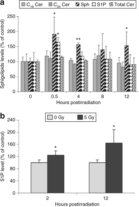
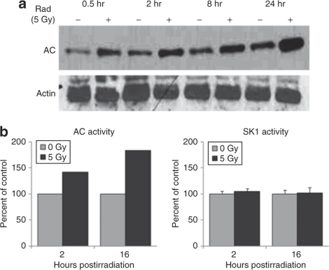
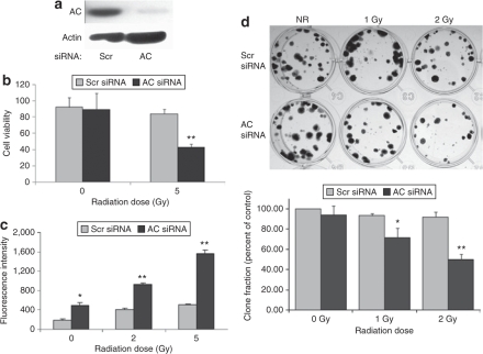
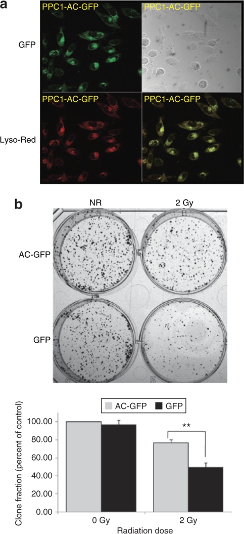
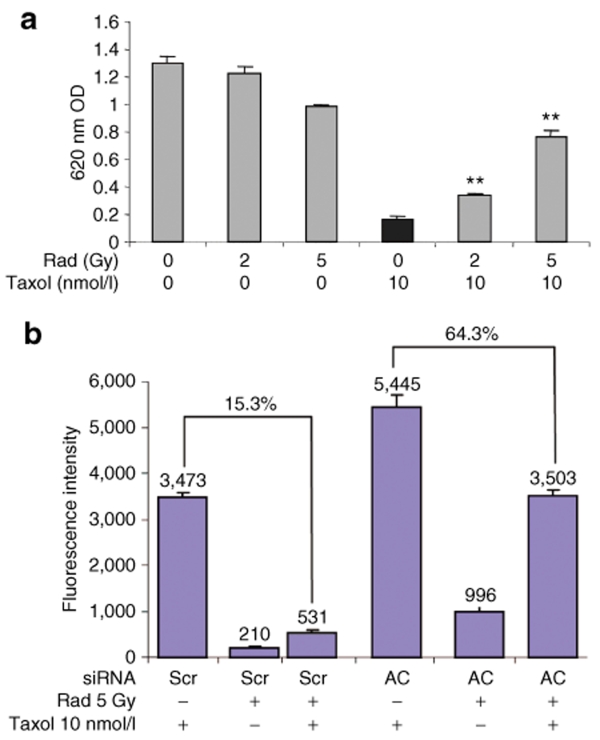
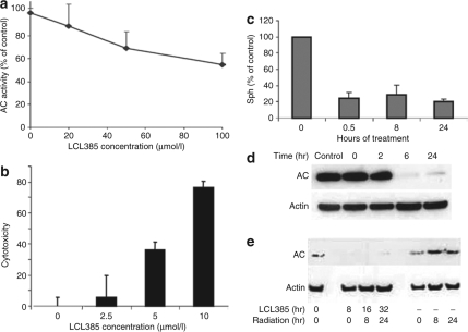
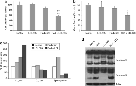
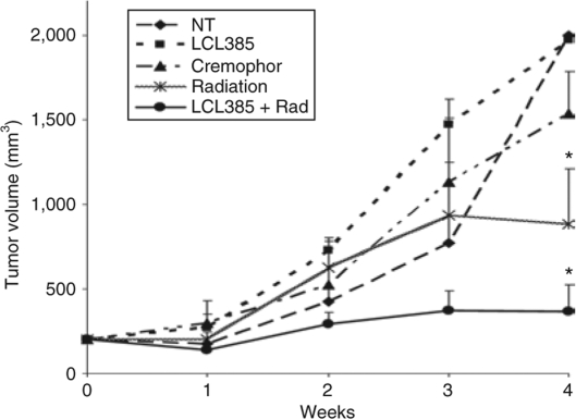
Radiation resistance in a subset of prostate tumors remains a challenge to prostate cancer radiotherapy. The current study on the effects of radiation on prostate cancer cells reveals that radiation programs an unpredicted resistance mechanism by upregulating acid ceramidase (AC). Irradiated cells demonstrated limited changes of ceramide levels while elevating levels of sphingosine and sphingosine-1-phosphate. By genetically downregulating AC with small interfering RNA (siRNA), we observed radiosensitization of cells using clonogenic and cytotoxicity assays. Conversely, AC overexpression further decreased sensitivity to radiation. We also observed that radiation-induced AC upregulation was sufficient to create cross-resistance to chemotherapy as demonstrated by decreased sensitivity to Taxol and C(6) ceramide compared to controls. Lower levels of caspase 3/7 activity were detected in cells pretreated with radiation, also indicating increased resistance. Finally, utilization of the small molecule AC inhibitor, LCL385, sensitized PPC-1 cells to radiation and significantly decreased tumor xenograft growth. These data suggest a new mechanism of cancer cell resistance to radiation, through upregulation of AC that is, in part, mediated by application of the therapy itself. An improved understanding of radiotherapy and the application of combination therapy achieved in this study offer new opportunities for the modulation of radiation effects in the treatment of cancer.


Figures

<b>Figure 1</b>
Figure 1
Sphingosine and sphingosine-1-phosphate (S1P), but not ceramide (Cer), are upregulated by ionizing radiation. PPC-1 prostate cancer cell cultures were irradiated (mock or 5 Gy) and collected at the indicated time points after irradiation. Cell pellets and culture media were prepared and lipids were extracted for mass spectrometry as mentioned in Materials and Methods. (a) Intracellular C16 Cer (shaded bars), C24 Cer (hatched bars), sphingosine (Sph, striped bars), S1P (diamond bars), and total Cer (dotted bars) expression levels are represented as percent of nonirradiated cells. (b) S1P levels in PPC-1 culture media from irradiated or control cells. Results shown are mean ± SD of three replicates from one of two independent, representative experiments. *P < 0.05, **P < 0.01 compared with nonirradiated cells.
<b>Figure 2</b>
Figure 2
Ionizing radiation induces upregulation of acid ceramidase (AC), but not sphingosine kinase-1 (SK1). PPC-1 prostate cancer cells were irradiated (5 Gy) and collected during the first 24 hours of irradiation. (a) Protein lysates were subjected to western blot analysis for AC protein expression. (b) Protein lysates were isolated at 2 and 16 hours following irradiation, and in vitro AC and SK1 enzymatic activities were evaluated as described in Materials and Methods.
<b>Figure 3</b>
Figure 3
Acid ceramidase (AC) silencing reverses the insensitivity of PPC-1 cells to ionizing radiation. PPC-1 cells were plated in 35-mm2 dishes at a concentration of 1 × 10⁵ cells/dish. Following overnight incubation, cells were transfected with small interfering RNA (siRNA) targeting either AC or a scrambled (Scr)-sequence control. (a) AC protein expression was evaluated by western blot 30 hours following siRNA transfection. (b) Twenty-four hours after siRNA transfection, cells were irradiated at 0 or 5 Gy, and cell death was monitored at 48 hours postirradiation by MTS assay. Cell viability data are represented as percent of untreated cells. (c) Caspase 3/7 activity was determined 24 hours after irradiation to measure the apoptotic status in irradiated or control cells. (d) Irradiated and control cells were seeded at 200 cells/well and cultured for 14 days. Cultures were then fixed and stained with crystal violet. Representative examples are presented. The bar graph below represents quantification by colony scoring. Results are representative of the average ± SD of three independent experiments, each performed as four replicates. *P < 0.05, **P < 0.01 compared with Scr-sequence-transfected cells.
<b>Figure 4</b>
Figure 4
Acid ceramidase (AC) over-expression reduces PPC-1 cell sensitivity to ionizing radiation. PPC-1 cells were transfected with plasmid constructs expressing either an AC-GFP fusion protein or green fluorescent protein (GFP) (control). Stably transfected clones were selected and validated for AC overexpression by western blot. (a) Confocal microscopic images of AC-GFP (green), LysoTracker Red–labeled lysosomes (red), or overlay of the two colors is shown and indicates the proper AC colocalization (yellow) within the lysosome. (b) AC overexpressing PPC-1 cells and control cells were irradiated and a clonogenic assay was performed as described in Materials and Methods. After 2 weeks in culture, cells were fixed and stained with crystal violet. Representative cell cultures are depicted. The bar graph below represents quantification by colony scoring. Results are presented as percent of nonirradiated control cells. Results are representative of the average ± SD of two independent experiments, each performed in triplicate. **P < 0.01 compared to GFP-transfected cells.
<b>Figure 5</b>
Figure 5
Radiation desensitizes PPC-1 cells to Taxol treatment by upregulation of acid ceramidase (AC). (a) PPC-1 cells were irradiated at 0, 2, or 5 Gy followed 2 hours later by treatment with 10 nmol/l Taxol. After 48 hours of chemotherapy, attached cells were fixed and evaluated by crystal violet staining intensity as measured by optical density (OD) at 620 nm. (b) PPC-1 cells were pretransfected with control or AC small interfering RNA (siRNA) 24 hours prior to radiation. Two hours after radiation, 10 nmol/l Taxol was applied. The caspase 3/7 activity assay was performed 24 hours after chemotherapy and results depicted as fluorescence intensity. Results are representative of the average ± SD of four independent experiments. Each data point is the average of three replicates. **P < 0.01 compared with Taxol treatment alone. Scr, scrambled.
<b>Figure 6</b>
Figure 6
Acid ceramidase (AC) inhibitor LCL385 blocks radiation-induced AC upregulation. (a) Dose-dependent inhibitory effect of LCL385 on AC in vitro was performed through a cell-free assay using PPC-1 cell homogenates as the AC source and [3H-N] labeled C6 ceramide as the substrate, performed at pH 4.5. Data are plotted as percentage of AC inhibition compared to control cell lysate. (b) Cytotoxicity of LCL385 on PPC-1 cells was evaluated by MTS assay 24 hours following treatment at the indicated doses. Results represent percentage of cell death compared to cells with no treatment. (c) PPC-1 cells were treated with LCL385 (2.5 µmol/l). At the indicated time points sphingosine levels were measured in triplicate using mass spectrometry and plotted as the percent change compared to cells with no LCL385 treatment. (d) PPC-1 cells were treated with LCL385 (2.5 µmol/l) and protein lysates were isolated at the indicated time points and processed by western blotting for AC protein expression. (e) PPC-1 cells were treated with LCL385 (2.5 µmol/l) 8 hours prior to radiation as indicated. Protein lysates were collected at 8 or 24 hours after radiation administration and processed by western blotting to determine AC protein levels.
<b>Figure 7</b>
Figure 7
Acid ceramidase (AC) inhibition sensitizes PPC-1 cells to ionizing radiation. PPC-1 cells were pretreated with LCL385 at 2.5 µmol/l followed 8 hours later by 5 Gy radiation. (a) Cell viability at 48 hours following irradiation was evaluated by crystal violet staining. Results are represented as percent of untreated control cells quantified by colorimetric absorbance at 620 nm. (b) Clonogenic survival between 10 and 14 days was evaluated as described in Materials and Methods. Results are presented as percent of nonirradiated crystal violet staining in control cells. (c) Sphingolipid species in treated cells at 30 minutes following radiation were analyzed by mass spectrometry. (d) Protein lysates were collected at 48 hours following irradiation and/or LCL385 treatment, and processed by western blot for assessment of caspases 3 and 9 activation. *P < 0.05; ** and ††P < 0.01 compared with radiation alone and LCL385 treatment alone, respectively.
<b>Figure 8</b>
Figure 8
Acid ceramidase (AC) inhibitor LCL385 sensitizes PPC-1 tumor xenografts to ionizing radiation. Eight million PPC-1 tumor cells were injected subcutaneously into the flank of nu/nu mice. When tumors reached a mean size of 200 mm3, animals received 5 Gy of radiation (Varian 2100c linear accelerator) twice weekly for a total of six doses (30 Gy total) alone or in combination with LCL385 administered by intraperitoneal injection at 30 mg/kg in 400 µl of Cremophor/ethanol/phosphate-buffered saline 8 hours prior to radiation as described in Materials and Methods. LCL385, no treatment, irradiation, or Cremophor alone were controls used for comparison. Tumor volume was measured twice weekly and expressed as mentioned in Materials and Methods. Data points represent the mean change in tumor volumes relative to day 0 after tumors reached ~200 mm3 for each group of animals (n = 6 or 7). *P < 0.05 compared to the control group.

References

    1. Mangar SA, Huddart RA, Parker CC, Dearnaley DP, Khoo VS., and , Horwich A. Technological advances in radiotherapy for the treatment of localised prostate cancer. Eur J Cancer. 2005;41:908–921. - PubMed
    1. Peschel RE., and , Colberg JW. Surgery, brachytherapy, and external-beam radiotherapy for early prostate cancer. Lancet Oncol. 2003;4:233–241. - PubMed
    1. Hanks GE, Hanlon AL, Epstein B., and , Horwitz EM. Dose response in prostate cancer with 8–12 years' follow-up. Int J Radiat Oncol Biol Phys. 2002;54:427–435. - PubMed
    1. Ward JF. DNA damage produced by ionizing radiation in mammalian cells: identities, mechanisms of formation, and reparability. Prog Nucleic Acid Res Mol Biol. 1988;35:95–125. - PubMed
    1. Allan DJ. Radiation-induced apoptosis: its role in a MADCaT (mitosis-apoptosis-differentiation-calcium toxicity) scheme of cytotoxicity mechanisms. Int J Radiat Biol. 1992;62:145–152. - PubMed

Publication types

MeSH terms