. 2008;3(12):e4026.
doi: 10.1371/journal.pone.0004026. Epub 2008 Dec 24.

HSP60 as a target of anti-ergotypic regulatory T cells

Affiliations

HSP60 as a target of anti-ergotypic regulatory T cells

Francisco J Quintana et al. PLoS One. 2008.

Abstract

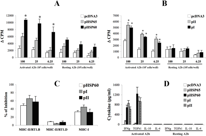
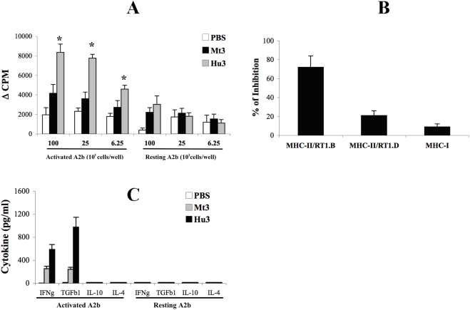
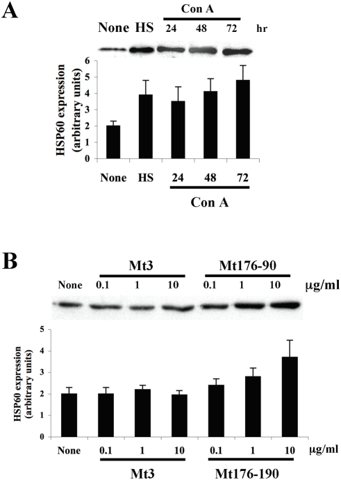
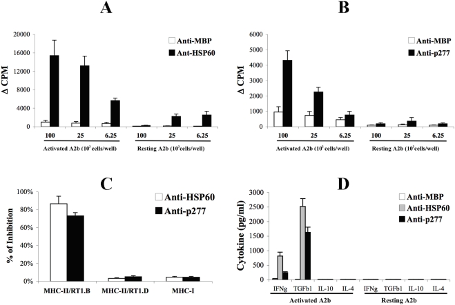
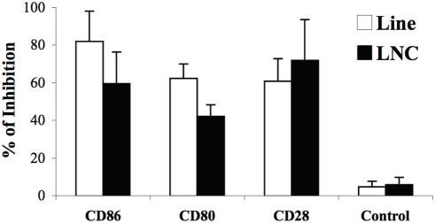
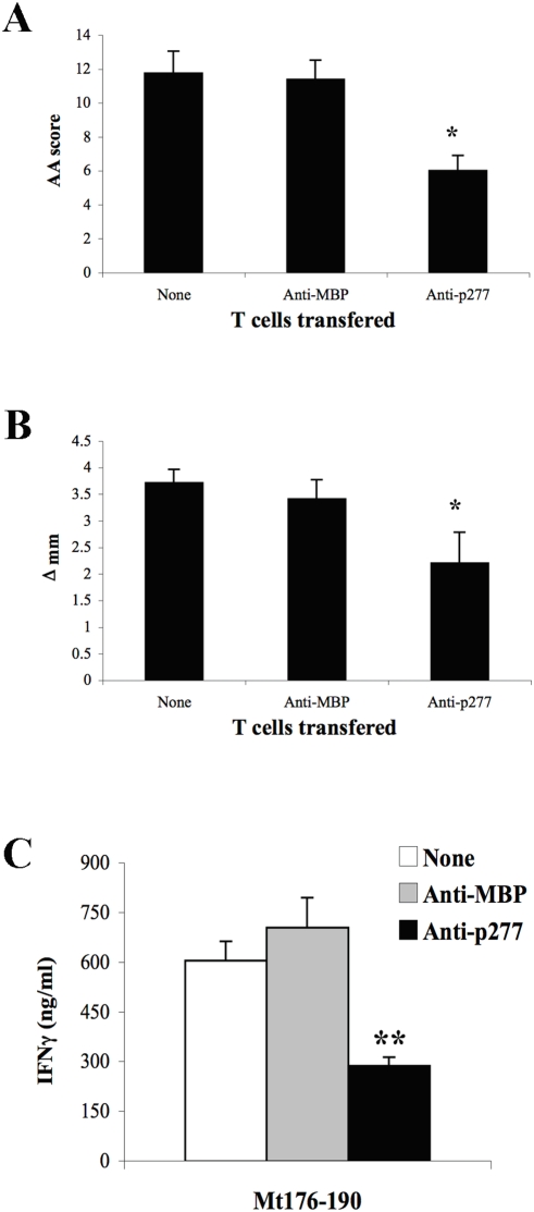
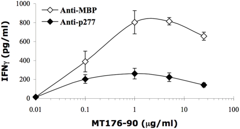
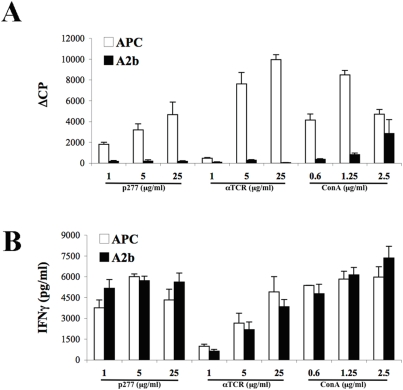
The 60 kDa heat shock protein (HSP60) has been reported to influence T-cell responses in two ways: as a ligand of toll-like receptor 2 signalling and as an antigen. Here we describe a new mechanism of T-cell immuno-regulation focused on HSP60: HSP60 is up-regulated and presented by activated T cells (HSP60 is an ergotope) to regulatory (anti-ergotypic) T cells. Presentation of HSP60 by activated T cells was found to be MHC-restricted and dependent on accessory molecules - CD28, CD80 and CD86. Anti-ergotypic T cells responded to T-cell HSP60 by proliferation and secreted IFNgamma and TGFbeta1. In vitro, the anti-ergotypic T cells inhibited IFNgamma production by their activated T-cell targets. In vivo, adoptive transfer of an anti-ergotypic HSP60-specific T-cell line led to decreased secretion of IFNgamma by arthritogenic T cells and ameliorated adjuvant arthritis (AA). Thus, the presentation of HSP60 by activated T cells turns them into targets for anti-ergotypic regulatory T cells specific for HSP60. However, the direct interaction between the anti-ergotypic T regulators (anti-HSP60) and the activated T cells also down-regulated the regulators. Thus, by functioning as an ergotope, HSP60 can control both the effector T cells and the regulatory HSP60-specific T cells that control them.


Conflict of interest statement

Competing Interests: The authors have declared that no competing interests exist.

Figures

Figure 1
Figure 1. DNA vaccination with HSP60 induces anti-ergotypic T cells.
A and B. Anti-ergotypic proliferative response of LNC from rats vaccinated with pcDNA3, pHSP65 or pHSP60 (A) or pcDNA3, pI or pII (B), taken 26 days after the induction of AA. Proliferative responses are presented as the ΔCPM±SEM of quadruplicate cultures. * p<0.05 compared to the pHSP65 (A) or the pcDNA3 (B) groups. C. Monoclonal antibodies to MHC-II/RT1.B, MHC-II/RT1.D or MHC-I were assayed for their ability to block the anti-ergotypic proliferative response. Results are presented as the percent of inhibition of proliferation±SEM of quadriplicate cultures. D. Anti-ergotypic cytokine response of LNC taken from rats vaccinated with pcDNA3, pHSP65, pHSP60, pI or pII 26 days after the induction of AA. IFNγ (IFNg), TGFβ1 (TGFb1), IL-10 and IL-4 were quantified in the culture supernatants after 72 hr of stimulation with 105 activated or resting, irradiated, A2b cells per well. The results are presented as pg/ml±SEM of triplicate cultures. Three independent experiments produced similar results.
Figure 2
Figure 2. Vaccination with HSP60 peptide Hu3 induces anti-ergotypic T cells.
A. Anti-ergotypic proliferative response of LNC from rats vaccinated with PBS, Mt3 or Hu3 in IFA, taken 26 days after AA induction. Proliferative responses are presented as the ΔCPM±SEM of quadruplicate cultures. * p<0.05 compared to the Mt3 group. B. Monoclonal antibodies to MHC-II/RT1.B, MHC-II/RT1.D or MHC-I were assayed for their ability to block the anti-ergotypic proliferative response. Results are presented as the percent of inhibition of proliferation±SEM of quadruplicate cultures. C. Anti-ergotypic cytokine response of LNC taken from rats vaccinated with PBS, Mt3 or Hu3 in IFA, 26 days after AA induction. IFNγ (IFNg), TGFβ1 (TGFb1), IL-10 and IL-4 were quantified in the culture supernatants after 72 hr of stimulation with 105 activated or resting, irradiated, A2b cells per well. The results are presented as pg/ml±SEM of triplicate cultures. Three independent experiments produced similar results.
Figure 3
Figure 3. T-cell activation up-regulates cellular levels of HSP60.
A. LNC were stimulated with Con A for 24, 48 or 72 hr, subjected to a 30 minutes 42°C heat shock (HS) or kept at 37°C (None). Cell lysates were prepared and HSP60 expression was analyzed by western blot with specific antibodies, and quantified (in arbitrary units). B. A2b T-cells were stimulated with various concentrations of the target peptide Mt176-90, a control peptide (Mt3) for 72 hr, or with medium alone (None). Cell lysates were prepared and HSP60 expression was analyzed by western blot with specific antibodies, and quantified (in arbitrary units). Two independent experiments produced similar results.
Figure 4
Figure 4. MHC class II-restricted recognition of activated T cells by HSP60-specific T-cells.
A. Anti-ergotypic proliferative response of Anti-HSP60 or Anti-MBP T cell lines. Proliferative responses are presented as the ΔCPM±SEM of quadruplicate cultures. B. Anti-ergotypic proliferative response of Anti-p277 or Anti-MBP T cell lines. Proliferative responses are presented as the ΔCPM±SEM of quadruplicate cultures. C. Monoclonal antibodies to MHC-II/RT1.B, MHC-II/RT1.D or MHC-I were assayed for their ability to block the anti-ergotypic proliferative response of the Anti-HSP60 and the Anti-p277 T cell lines. Results are presented as the percent of inhibition of proliferation±SEM of quadruplicate cultures. D. IFNγ (IFNg), TGFβ1 (TGFb1), IL-10 and IL-4 were quantified in the culture supernatants after 72 hr of stimulation of the Anti-MBP, Anti-p277 or Anti-HSP60 T cell lines with 105 activated or resting, irradiated, A2b cells per well. The results are presented as pg/ml±SEM of triplicate cultures. Three to five independent experiments produced similar results.
Figure 5
Figure 5. The activation HSP60-specific anti-ergotypic T cells requires co-stimulation.
Monoclonal antibodies to CD28, CD80 or CD86, or a control IgG (Control), were assayed for their ability to block the anti-ergotypic proliferative response of Anti-HSP60 T-cells (Line) or of LNC prepared from pHSP60-vaccinated rats (LNC). Results are presented as the percent of inhibition of proliferation±SEM of quadruplicate cultures. Three independent experiments produced similar results.
Figure 6
Figure 6. HSP60-specific anti-ergotypic T-cells control arthritogenic T-cells in vivo.
A and B. Anti-MBP or Anti-p277 T cells were injected ip into naïve Lewis rats and three days later AA was induced. Twenty-six days after AA induction, at the peak of AA, the AA clinical score (A) and the hind paw diameter (B) were determined. The bars represent the mean values ± SEM for each group of 8 rats. C. LNC were collected on day 26 after AA induction and the secretion of IFNγ upon stimulation with Mt176-90 was studied. The results are presented as pg/ml±SEM of triplicate cultures. Three independent experiments produced similar results. * p<0.05 and ** p<0.005 compared to the Anti-MBP group.
Figure 7
Figure 7. HSP60-specific Anti-ergotypic T-cells control arthritogenic T-cells in vitro.
LNC from Mt immunized rats (2.5×10⁵ per well) were activated with Mt176-90 for 72 hr in the presence of Anti-p277 or Anti-MBP T-cells (5×10⁴ per well). The secretion of IFNγ was determined by ELISA, the results are presented as pg/ml±SEM of triplicate cultures. The differences between the groups were significant (p<0.05) for antigen concentrations higher than 0.1 µg/ml. Three independent experiments produced similar results.
Figure 8
Figure 8. Anti-ergotypic HSP60-specific T cells become anergic after interacting with activated T cells.
Anti-ergotypic Anti-p277 T cells were stimulated for 3 days with irradiated, activated A2b cells (A2b) or with irradiated APC fed with the p277 peptide (APC). The Anti-p277 T cells were maintained for 4 additional days in culture, and stimulated with APC and p277 peptide, Con A or immobilized anti-TCR (αTCR) antibodies. T-cell proliferation (A) and IFNγ (B) release were measured after 3 days. The proliferative responses are presented as the ΔCPM (±SEM) (A), and the IFNγ as pg/ml±SEM (B) of triplicate cultures. Three to five independent experiments produced similar results.

References

    1. Elias D, Markovits D, Reshef T, van der Zee R, Cohen IR. Induction and therapy of autoimmune diabetes in the non-obese diabetic (NOD/Lt) mouse by a 65-kDa heat shock protein. Proc Natl Acad Sci U S A. 1990;87:1576–1580. - PMC - PubMed
    1. Elias D, Reshef T, Birk OS, van der Zee R, Walker MD, et al. Vaccination against autoimmune mouse diabetes with a T-cell epitope of the human 65-kDa heat shock protein. Proc Natl Acad Sci U S A. 1991;88:3088–3091. - PMC - PubMed
    1. Quintana FJ, Carmi P, Cohen IR. DNA vaccination with heat shock protein 60 inhibits cyclophosphamide-accelerated diabetes. J Immunol. 2002;169:6030–6035. - PubMed
    1. Elias D, Cohen IR. The hsp60 peptide p277 arrests the autoimmune diabetes induced by the toxin streptozotocin. Diabetes. 1996;45:1168–1172. - PubMed
    1. Lopez-Guerrero JA, Lopez-Bote JP, Ortiz MA, Gupta RS, Paez E, et al. Modulation of adjuvant arthritis in Lewis rats by recombinant vaccinia virus expressing the human 60-kilodalton heat shock protein. Infect Immun. 1993;61:4225–4231. - PMC - PubMed

Publication types

MeSH terms