. 2009 Jan;11(1):48-56.
doi: 10.1593/neo.81036.

Evaluation of angiogenesis using micro-computed tomography in a xenograft mouse model of lung cancer

Affiliations
Free PMC article

Evaluation of angiogenesis using micro-computed tomography in a xenograft mouse model of lung cancer

Rajkumar Savai et al. Neoplasia. 2009 Jan.
Free PMC article

Abstract

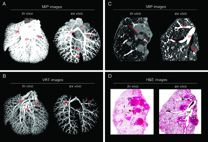
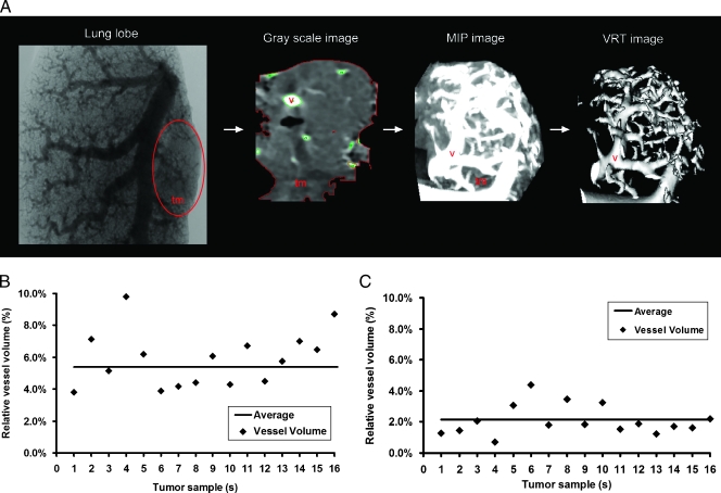
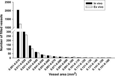
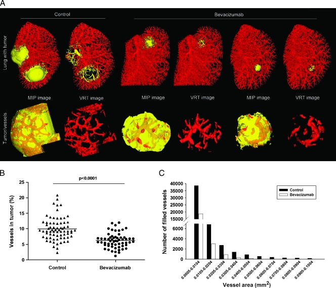
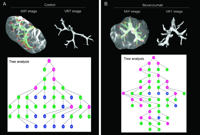
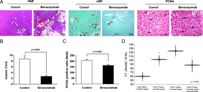
Quantitative evaluation of lung tumor angiogenesis using immunohistochemical techniques has been limited by difficulties in generating reproducible data. To analyze intrapulmonary tumor angiogenesis, we used high-resolution micro-computed tomography (micro-CT) of lung tumors of mice inoculated with mouse Lewis lung carcinoma (LLC1) or human adenocarcinoma (A549) cell lines. The lung vasculature was filled with the radiopaque silicone rubber, Microfil, through the jugular vein (in vivo application) or pulmonary artery (ex vivo application). In addition, human adenocarcinoma lung tumor-bearing mice treated site-specifically with humanized monoclonal antibody (bevacizumab) against vascular endothelial growth factor. Quantitative analysis of lung tumor microvessels imaged with micro-CT showed that more vessels (mainly small, <0.02 mm(2)) were filled using the in vivo (5.4%) compared with the ex vivo (2.1%) method. Furthermore, bevacizumab-treated lung tumor-bearing mice showed significantly reduced lung tumor volume and lung tumor angiogenesis compared with untreated mice as assessed by micro-CT. Interestingly, microvascularization of mainly the smaller vessels (<0.02 mm(2)) was reduced after bevacizumab treatment. This observation with micro-CT was nicely correlated with immunohistochemical measurement of microvessels. Therefore, micro-CT is a novel method for investigating lung tumor angiogenesis, and this might be considered as an additional complementary tool for precise quantification of angiogenesis.


Figures

Figure 1
Figure 1
Micro-CT and histology of an entire murine lung after in vivo or ex vivo injection of Microfil. (A) Three-dimensional MIP images of the entire lung after in vivo or ex vivo Microfil application. (B) Three-dimensional VRT images of the entire lung after in vivo or ex vivo Microfil application. Representative micro-CT and hematoxylin and eosin-stained images clearly showed the course of anatomical structures. (C) Three-dimensional MIP images (in vivo or ex vivo Microfil application) reconstructed from micro-CT images and the corresponding (D) histologic images (in vivo or ex vivo Microfil application). b indicates bronchus; es, esophagus; il, inflated lung; m, mediasternum; pa, pulmonary arteries; pv, pulmonary veins; tm, tumor mass; tr, trachea.
Figure 2
Figure 2
Quantitative volumetric measurements of extracted tumors and tumor angiogenesis after in vivo or ex vivo vessel filling. (A) A section of a Microfil-injected lung lobe: grayscale image of an extracted tumor from a Microfil-injected lung, MIP image showing a lung tumor, and a VRT image illustrating the tumor vasculature in a sequential order. Quantitative scatter graphs showing the percentage of filled vessels in tumors after in vivo (B) or ex vivo (C) Microfil application. tm indicates tumor mass; v, vessels (n = 16 tumors were taken for this analysis from 12 mice).
Figure 3
Figure 3
Quantitative measurement of tumor angiogenesis after in vivo or ex vivo vessel filling: bar graph showing the detailed information that vessels were in tumor by vessel area (mm2; n = 16 tumors were taken for this analysis from 12 mice).
Figure 4
Figure 4
Measurements of tumor angiogenesis after site-specific therapy. (A) Three-dimensional surface rendering images of lung tumors from untreated and bevacizumab-treated mice. Extracted tumors and tumor microvascularization are shown (yellow represents tumor tissue area; red represents tumor vessel area). (B) Number of vessels measured with micro-CT after 14 days of bevacizumab treatment (•) or without treatment (▴). (C) Bar graph showing the detailed information that vessels were in tumor by vessel area (mm2; n = 12 mice for each group; extracted tumors from each mouse = ∼5–6).
Figure 5
Figure 5
Lung tumor vascular branching by tree analysis: 3D MIP images, 3D VRT images, and vessel branching micrographs acquired from tree analysis of micro-CT of tumors from untreated (A) and bevacizumab-treated (B) mice. Colored circles in the branching schematics show the vessel branch points in the tumor and the vessel area: blue circles, <25 mm2; green circles, 25 to 100 mm2; pink circles, 100 to 150 mm2 (n = 6 tumors were used for each group).
Figure 6
Figure 6
Histopathology of lung tumors that received bevacizumab therapy. (A) Histologic images of sections (3 µm) from untreated and bevacizumab-treated lung tumor sections that were stained with hematoxylin and eosin (H&E; the black dashed lines indicate areas of hemorrhage in the tumor), with anti-vWF (red arrows in A indicate vWF-positive vessels) and anti-PCNA (black arrows in A indicate PCNA-positive cells), respectively. Quantification of vWF-positive tumor vessels (B) and PCNA-positive tumor cells (C) by counting positively stained cells in ten randomly selected microscopic fields in seven mice is given. (D) Lung and lung tumor tissue density with or without Microfil were measured with micro-CT (60 areas were selected). Means ± SEM are shown. Scale bars, 40 µm.

References

    1. Jemal A, Siegel R, Ward E, Hao Y, Xu J, Murray T, Thun MJ. Cancer statistics, 2008. CA Cancer J Clin. 2008;58:71–96. - PubMed
    1. Quinn MJ. Cancer trends in the United States—a view from Europe. J Natl Cancer Inst. 2003;95:1258–1261. - PubMed
    1. Folkman J. What is the evidence that tumors are angiogenesis dependent? J Natl Cancer Inst. 1990;82:4–6. - PubMed
    1. Conway EM, Collen D, Carmeliet P. Molecular mechanisms of blood vessel growth. Cardiovasc Res. 2001;49:507–521. - PubMed
    1. Folkman J. Angiogenesis: an organizing principle for drug discovery? Nat Rev Drug Discov. 2007;6:273–286. - PubMed

MeSH terms

LinkOut - more resources