

Quality of bronchial biopsies for morphology study and cell sampling: a comparison of asthmatic and healthy subjects
- PMID: 19107244
- PMCID: PMC2682166
- DOI: 10.1155/2008/202615
Quality of bronchial biopsies for morphology study and cell sampling: a comparison of asthmatic and healthy subjects
Abstract
Background: Bronchial biopsies are widely used for histopathological, primary cell culture and genetic studies, but very few reports have evaluated their quality.
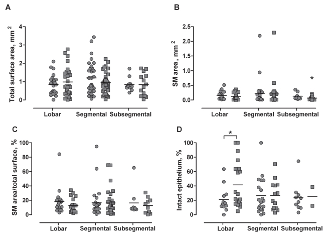
Objectives and methods: The present project evaluated the quality (using a scoring system) and the general morphology of a pool of six bronchial biopsy specimens taken from three different sampling sites (the lobar, segmental and subsegmental carinae) in 27 subjects (13 asthmatic subjects and 14 healthy controls). The present study also assessed quantitative measurements of structural changes related to asthma.
Results: In total, 94.4% of the biopsy attempts had enough tissue to be processed. From these, 61.7% were scored with a good to excellent quality, while 76.5% presented smooth muscle bundles and 40.5% had an intact epithelium wall. The data also confirmed the structural changes observed in asthma, such as increased apparent thickening of the basement membrane, reduced amounts of smooth muscle for healthy controls and decreased percentage of intact epithelium for asthmatic subjects.
Conclusion: A pool of six bronchial biopsy specimens can provide tissue of excellent quality in both asthmatic and healthy subjects and, consequently, a valuable sample for morphological analysis of mucosal structures.
HISTORIQUE :: Les biopsies bronchiques sont généralisées pour les études histopathologiques, génétiques et de cellules primaires, mais très peu de rapports en évaluent la qualité.
OBJECTIFS ET MÉTHODOLOGIE :: Le présent projet visait à évaluer la qualité (au moyen d’un système de pointage) et la morphologie générale d’un groupe de six spécimens de biopsie bronchique prélevés dans divers sites de prélèvements (les carènes lobaire, segmentaire et sous-segmentaire) de 27 sujets (13 sujets asthmatiques et 14 sujets témoins). La présente étude a également servi à évaluer des mesures quantitatives de changements structurels reliés à l’asthme.
RÉSULTATS :: Au total, 94,4 % des tentatives de biopsie contenaient assez de tissus pour être traitées. De ce nombre, 61,7 % obtenaient une évaluation de bonne à excellente qualité, tandis que 76,5 % comportaient des faisceaux de muscles lisses et 40,5 %, une paroi épithéliale intacte. Les données ont également confirmé les changements structurels observés dans l’asthme, tels qu’une augmentation de l’épaississement apparent de la couche basale, une moins grande quantité de muscle lisse chez les sujets en santé et une diminution du pourcentage d’épithélium intact chez les sujets asthmatiques.
CONCLUSIONS :: Un groupe de six spécimens de biopsie bronchique peut fournir des tissus d’excellente qualité tant chez les sujets asthmatiques que chez les sujets en santé et, par conséquent, un échantillon utile d’analyse morphologique des structures muqueuses.
Figures
References
-
- Elston WJ, Whittaker AJ, Khan LN, et al. Safety of research bronchoscopy, biopsy and bronchoalveolar lavage in asthma. Eur Respir J. 2004;24:375–7. - PubMed
-
- Azzawi M, Bradley B, Jeffery PK, et al. Identification of activated T lymphocytes and eosinophils in bronchial biopsies in stable atopic asthma. Am Rev Respir Dis. 1990;142:1407–13. - PubMed
-
- Beasley R, Roche WR, Roberts JA, Holgate ST. Cellular events in the bronchi in mild asthma and after bronchial provocation. Am Rev Respir Dis. 1989;139:806–17. - PubMed
-
- Jeffery P, Holgate S, Wenzel S. Methods for the assessment of endobronchial biopsies in clinical research: Application to studies of pathogenesis and the effects of treatment. Am J Respir Crit Care Med. 2003;168:S1–17. - PubMed
Publication types
MeSH terms
LinkOut - more resources
Full Text Sources
Medical
