

Is a preserved functional reserve a mechanism limiting clinical impairment in pediatric MS patients?
- PMID: 19107755
- PMCID: PMC6871244
- DOI: 10.1002/hbm.20712
Is a preserved functional reserve a mechanism limiting clinical impairment in pediatric MS patients?
Abstract
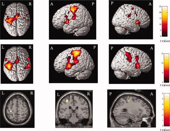
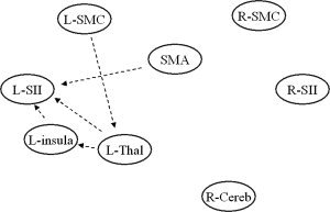
We evaluated the functional magnetic resonance imaging (fMRI) correlates of simple movement performance in patients with pediatric multiple sclerosis (MS) and their relation with the extent of T2 lesion volume (LV), to improve our understanding of the mechanisms leading to their short/medium term favorable clinical course. We obtained fMRI during repetitive flexion-extension of the last four fingers of the right hand and brain dual-echo scans from 17 right-handed patients with pediatric relapsing-remitting MS and 9 sex- and age-matched right-handed healthy controls. T2 LV was measured using a local thresholding segmentation technique. fMRI activations and functional connectivity analysis were performed using SPM2. Compared to controls, pediatric MS patients had an increased recruitment of the left (L) primary sensorimotor cortex (SMC). They also showed reduced functional connectivity between the L primary SMC and the L thalamus (P = 0.03), the L insula and the L secondary sensorimotor cortex (SII) (P = 0.02), the supplementary motor area and the L SII (P = 0.02), the L thalamus and the L insula (P = 0.01) and the L thalamus and the L SII (P = 0.003). In patients with pediatric MS, the activity of the L primary SMC was significantly correlated with brain T2 LV (r = 0.78). No correlation was found between coefficients of abnormal connectivity and structural MRI measures. The maintenance of a selective and strictly lateralized pattern of movement-associated brain activations and a modulation of its functional connections suggest a preserved functional reserve in patients with pediatric MS, which, in turn, might contribute to explain their favorable clinical evolution at short/medium term.
2008 Wiley-Liss, Inc.
Figures
References
-
- Balassy C,Bernert G,Wöber‐Bingöl C,Csapó B,Kornek B,Széles J,Fleischmann D,Prayer D ( 2001): Long‐term MRI observations of childhood‐onset relapsing‐remitting multiple sclerosis. Neuropediatrics 32: 28–37. - PubMed
-
- Banwell B,Ghezzi A Bar‐Or A, Mikaeloff Y,Tardieux M ( 2007a): Multiple sclerosis in children: Clinical diagnosis, therapeutic strategies, and future directions. Lancet Neurol 6: 887–902 (Review). - PubMed
-
- Banwell B,Shroff M,Ness JM,Jeffery D,Schwid S,Weinstock‐Guttman B,International Pediatric MS Study Group ( 2007b): MRI features of pediatric multiple sclerosis. Neurology 68: S46–S53. - PubMed
-
- Calautti C,Baron JC ( 2003): Functional neuroimaging studies of motor recovery after stroke in adults: A review. Stroke 34: 1553–1566. - PubMed
Publication types
MeSH terms
LinkOut - more resources
Full Text Sources
Medical
