Comparative Study
. 2008 Dec;64(6):654-63.
doi: 10.1002/ana.21511.

Glucose and NADPH oxidase drive neuronal superoxide formation in stroke

Affiliations
Comparative Study

Glucose and NADPH oxidase drive neuronal superoxide formation in stroke

Sang Won Suh et al. Ann Neurol. 2008 Dec.

Abstract

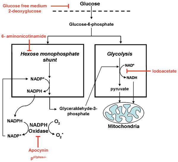
Objective: Hyperglycemia has been recognized for decades to be an exacerbating factor in ischemic stroke, but the mechanism of this effect remains unresolved. Here, we evaluated superoxide production by neuronal nicotinamide adenine dinucleotide phosphate (NADPH) oxidase as a possible link between glucose metabolism and neuronal death in ischemia-reperfusion.

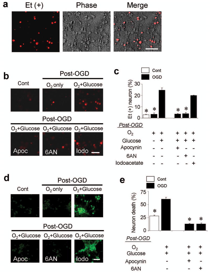
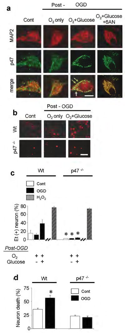
Methods: Superoxide production was measured by the ethidium method in cultured neurons treated with oxygen-glucose deprivation and in mice treated with forebrain ischemia-reperfusion. The role of NADPH oxidase was examined using genetic disruption of its p47(phox) subunit and with the pharmacological inhibitor apocynin.

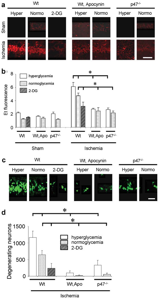
Results: In neuron cultures, postischemic superoxide production and cell death were completely prevented by removing glucose from the medium, by inactivating NADPH oxidase, or by inhibiting the hexose monophosphate shunt that generates NADPH from glucose. In murine stroke, neuronal superoxide production and death were decreased by the glucose antimetabolite 2-deoxyglucose and increased by high blood glucose concentrations. Inactivating NADPH oxidase with either apocynin or deletion of the p47(phox) subunit blocked neuronal superoxide production and negated the deleterious effects of hyperglycemia.

Interpretation: These findings identify glucose as the requisite electron donor for reperfusion-induced neuronal superoxide production and establish a previously unrecognized mechanism by which hyperglycemia can exacerbate ischemic brain injury.


Conflict of interest statement

Conflict of interest statement: None of the authors have any financial interest relevant to the work presented in this manuscript.

Figures

Figure 1
Figure 1. Metabolic coupling between glucose and superoxide production
Glucose can support superoxide production by supplying reducing equivalents to either NADPH oxidase or the mitochondria. Glucose transport and entry into cells is blocked by glucose-free medium or by 2-deoxyglucose. Flux of glucose carbon and glucose-derived NADPH to mitochondria is blocked by iodoacetate at the glyceraldehyde-3-phosphate dehydrogenase step of glycolysis. The production of NADPH in the hexose monophosphate shunt is blocked by 6-aminonicotinamide, and the activity of NADPH oxidase is blocked by apocynin or by p47phox deficiency.
Figure 2
Figure 2. Glucose is required for neuronal superoxide production after oxygen - glucose deprivation (OGD)
(a) Neuronal superoxide production imaged by ethidium (Et) fluorescence. Cultures underwent 2 hours of OGD followed by 1 hour incubation in air-equilibrated medium containing 5 mM glucose (O2 + glucose). Ethidium fluorescence is evident in a subset of the neuronal cell bodies. Scale bar = 100 μm. (b) Et fluorescence was markedly reduced in cultures maintained in glucose-free (O2 only) medium for the 1-hour interval after OGD. Control wells received medium exchanges only. Where indicated, cultures were also treated with apocynin (Apoc, 500 μM), 6-aminonocotinamide (6AN, 500 μM) or iodoacetate (Iodo, 500 μM). (c) Quantified ethidium fluorescence, conditions as in (b). The increased signal observed after OGD was prevented by 0-glucose medium, apocynin, and 6-aminonicotinamide, but not by iodoacetate. n = 7, * P < 0.05 vs. OGD, O2+glucose. (d) Immunostaining for 4-hydroxynonenal in cultured neurons showed the same pattern as observed with ethidium fluorescence. Conditions as in (b); representative of n = 3. Scale bar = 100 μm. (e) Neuronal death in cultures treated as in (b), assessed 24 hours after OGD. n = 3-5, * P < 0.05 vs. OGD, O2+glucose.
Figure 3
Figure 3. p47phox -/- neurons do not produce superoxide after OGD
(a) In wild-type neurons, immunostaining for the p47phox subunit of NADPH oxidase shows migration to the neuronal plasma membrane area (arrows) in neurons placed in standard medium after OGD, but not in neurons placed in 0-glucose medium or treated with 6-aminonicotinamide (500 μM) after OGD. MAP2 immunostaining demarcates the neuronal cytoplasmic area. Scale bar = 10 μm. Representative of n = 3. (b) Ethidium fluorescence in wild-type (wt) and p47phox -/- neurons subjected to 2 hours OGD, followed by 1 hour incubation in standard medium (O2 + glucose) or glucose-free medium (O2 only). Scale bar = 100 μm. (c) Quantified ethidium fluorescence. H2O2, 30 minutes exposure to 100 μM hydrogen peroxide; other conditions as in (a). n = 3-8, * P < 0.05 vs. wt neurons treated identically. (d) OGD - reperfusion produced less death in p47phox -/- neurons than wt neurons. n = 5, *P < 0.05 vs. control.
Figure 4
Figure 4. Ischemia-induced neuronal superoxide production and cell death are influenced by blood glucose and NADPH oxidase activity
(a) Ethidium fluorescence in CA1 hippocampal neurons 3 hours after ischemia-reperfusion. Sham mice received surgery without ischemia. Mice were wild-type (wt) or p47phox-/- genotype. Apo, 15 mg / kg apocynin; 2-DG, 100 mg / kg 2-deoxyglucose. Scale bar = 100 μm. (b) Quantification of ethidium fluorescence, conditions as in (a). n = 4- 5, * P < 0.05. (c) Fluoro-Jade-B staining of degenerating neurons in CA1 hippocampus 3 days after ischemia. (d) Quantified neuronal death; n = 7 - 10, * P < 0.05.

References

    1. National Institute of Neurological Disorders and Stroke rt-PA Stroke Study Group. Tissue plasminogen activator for acute ischemic stroke. The National Institute of Neurological Disorders and Stroke rt-PA Stroke Study Group. N Engl J Med. 1995;333:1581–1587. - PubMed
    1. Zivin JA, Lyden PD, DeGirolami U, et al. Tissue plasminogen activator. Reduction of neurologic damage after experimental embolic stroke. Arch Neurol. 1988;45:387–391. - PubMed
    1. Aronowski J, Strong R, Grotta JC. Reperfusion injury: demonstration of brain damage produced by reperfusion after transient focal ischemia in rats. J Cereb Blood Flow Metab. 1997;17:1048–1056. - PubMed
    1. Hallenbeck JM, Dutka AJ. Background review and current concepts of reperfusion injury. Arch Neurol. 1990;47:1245–1254. - PubMed
    1. Martini SR, Kent TA. Hyperglycemia in acute ischemic stroke: a vascular perspective. J Cereb Blood Flow Metab. 2007;27:435–451. - PubMed

Publication types