. 2008 Dec 31;49(6):1023-31.
doi: 10.3349/ymj.2008.49.6.1023.

Development of monoclonal antibodies against human IRF-5 and their use in identifying the binding of IRF-5 to nuclear import proteins karyopherin-alpha1 and -beta1

Affiliations

Development of monoclonal antibodies against human IRF-5 and their use in identifying the binding of IRF-5 to nuclear import proteins karyopherin-alpha1 and -beta1

Soo-In Yeon et al. Yonsei Med J. .

Abstract

Purpose: IRF-5 is a direct transducer of virus-mediated and TLR-mediated signaling pathways for the expression of cytokines and chemokines which form homodimers or heterodimers with IRF-7. However, direct IRF-5-specific monoclonal antibodies (mAbs) are not available at present. These could be used to further evaluate the functions of IRF-5. In this study, we produced and characterized three mouse mAbs to human IRF-5. The binding of IRF-5 to nuclear import proteins was first identified using a mAb.

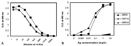
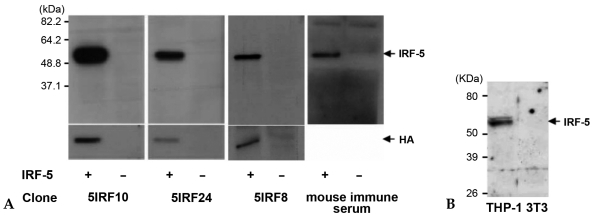
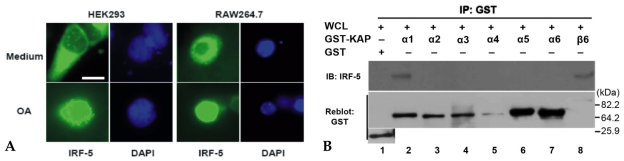
Materials and methods: His-tagged human IRF-5 protein spanning amino acid residues 193-257 was used as an antigen and three mAbs were produced. The mAbs were tested with ELISA, Western blot analysis (WB), immunofluorescent staining (IF), and immunoprecipitation (IP). In addition, the nuclear import protein which carried phosphorylated IRF-5 was identified using one of these mAbs.

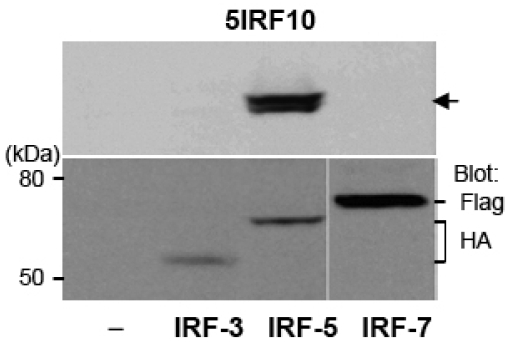
Results: MAbs 5IRF8, 5IRF10 and 5IRF24 which reacted with the recombinant His-IRF-5(193-257) protein were produced. All mAbs bound to human IRF-5, but not to IRF-3 or IRF-7. They could be used for WB, IF, and IP studies. The binding of phosphorylated IRF-5 to karyopherin-alpha1 and -beta1 was also identified.

Conclusion: Human IRF-5-specific mAbs are produced for studying the immunologic roles related to IRF-5. Phosphorylated IRF-5 is transported to the nucleus by binding to nuclear import proteins karyopherin-alpha1 and -beta1.


Figures

Fig. 1
Fig. 1
SDS-PAGE and MALDI-TOF mass spectrum of recombinant His-IRF-5193-257. (A) SDS-PAGE analysis of purified His-IRF-5193-257. His-IRF-5193-257 was eluted with 200 mM imidazole on a Ni++-NTA agarose resin and purifed by Sephacryl S-200 gel chromatography. (B) MALDI-TOF mass spectrum analysis of purified His-IRF-5193-257 protein. The molecular weight (MW) was determined to be 10728.36 Da using MALDI-TOF mass spectrum. This was very close to the theoretical MW of 10721.1 Da.
Fig. 2
Fig. 2
Binding curve between the anti-IRF-5 monoclonal antibodies and the His-IRF-5193-257 protein. (A) ELISA was performed to the wells. The wells were coated with 1 µg/mL of His-IRF-5193-257 protein using the various dilutions of culture supernatants of our three mAbs. (B) ELISA was performed with mAbs at a fixed dilution to the wells which were coated with various concentrations of the His-IRF-5193-257 protein. The bindings of all three mAbs to the control protein of α-synuclein are shown inside the black triangle.
Fig. 3
Fig. 3
Western blot analysis for antigen specificity. (A) An HA-tagged IRF-5 plasmid was transfected into HEK293 cells and the WCL was separated. The reactivity of mAbs to IRF-5 was tested. The expression of IRF-5 was tested with anti-HA. Mouse immune serum was used as a positive control Ab. (B) Detection of endogenous IRF-5. WCLs of THP-1 and NIH3T3 cells were separated and immunoblotted with 5IRF10 as a representative study. NIH3T3 cell lysate was used as a negative control.
Fig. 4
Fig. 4
Cross-reactive binding study. HA-tagged IRF-3, HA-tagged IRF-5, and Flag-tagged IRF-7 plasmids were transfected into HEK293 cells. WCLs were separated and the cross-reactivity of 5IRF10 to IRF-3 and IRF-7 was tested as a representative study. The membranes were reblotted with anti-HA or anti-Flag depending on the tagged protein. Arrow: IRF-5.
Fig. 5
Fig. 5
Immunofluorescence staining of IRF-5. HEK293 cells were transfected with a GFP-tagged IRF-5 plasmid. The cells were fixed and stained with the indicated mAbs for the His-IRF-5193-257 protein. PE-labeded anti-mouse Ig was added to the cells. The merged images are shown. Bar: 10 µm.
Fig. 6
Fig. 6
Immunoprecipitation. (A) HEK293 cells were transfected with a GFP-tagged IRF-5 plasmid. The WCL was immunoprecipitated with 5IRF10. The bound proteins were separated and the membrane was blotted with anti-GFP. WCLs were used as controls (lanes 1 and 3). (B) Immunoprecipitation of endogenous IRF-5 protein. WCLs of human cell lines of THP-1 and HEK293 were immunoprecipitated with 5IRF10. They were separated and blotted with 5IRF24. HEK293 cell lysate was used as a negative control.
Fig. 7
Fig. 7
Import of phosphorylated IRF-5 to the nucleus. (A) The localization of phosphorylated IRF-5 to the nucleus. HEK293T cells and RAW264.7 cells were transfected with an IRF-5 plasmid and treated with 20 nM OA for 6 h after transfection. The cells were stained with 5IRF10, followed by FITC-conjugated anti-mouse Ig. DAPI was used for staining the nucleus. Bar: 10 µm. (B) The binding of IRF-5 to KAP-α1 and -β1. GST-KAPs-α1, -α2, -α3, -α4, -α5, -α6 and -β1 fusion proteins immobilized on glutathione-Sepharose 4B beads were incubated overnight at 4℃ with WCLs of HEK293 cells transfected with an IRF-5 plasmid. Sepharose-bound proteins were separated and the membrane was blotted with 5IRF10. It was then reblotted with anti-GST.

References

    1. Miyamoto M, Fujita T, Kimura Y, Maruyama M, Harada H, Sudo Y, et al. Regulated expression of a gene encoding a nuclear factor, IRF-1, that specifically binds to IFN-beta gene regulatory elements. Cell. 1988;54:903–913. - PubMed
    1. Lohoff M, Mak TW. Roles of interferon-regulatory factors in T-helper-cell differentiation. Nat Rev Immunol. 2005;5:125–135. - PubMed
    1. Taniguchi T, Ogasawara K, Takaoka A, Tanaka N. IRF family of transcription factors as regulators of host defense. Annu Rev Immunol. 2001;19:623–655. - PubMed
    1. Negishi H, Ohba Y, Yanai H, Takaoka A, Honma K, Yui K, et al. Negative regulation of Toll-like-receptor signaling by IRF-4. Proc Natl Acad Sci U S A. 2005;102:15989–15994. - PMC - PubMed
    1. Sato M, Suemori H, Hata N, Asagiri M, Ogasawara K, Nakao K, et al. Distinct and essential roles of transcription factors IRF-3 and IRF-7 in response to viruses for IFN-alpha/beta gene induction. Immunity. 2000;13:539–548. - PubMed

Publication types