

Pleural epithelioid hemangioendothelioma
- PMID: 19108030
- PMCID: PMC2628035
- DOI: 10.3349/ymj.2008.49.6.1036
Pleural epithelioid hemangioendothelioma
Abstract
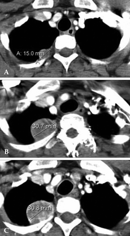
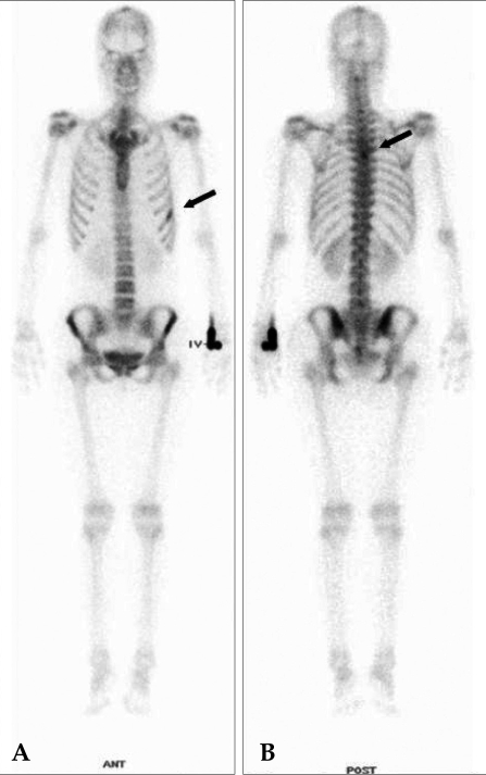
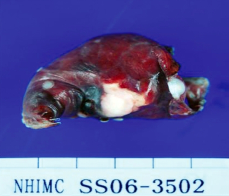
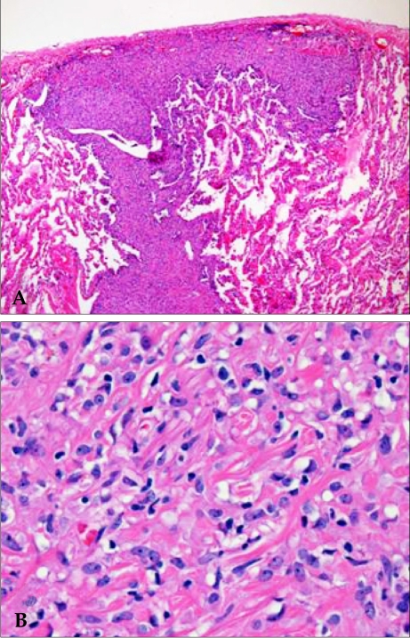
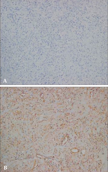
Epithelioid hemangioendothelioma (EHE) is a rare tumor of vascular origin. While it can be found in any tissue, it is most often found in lung and liver and usually has an intermediate behavior. EHEs originating from pleural tissue have been less frequently described than those from other sites. Furthermore, to date, all of the cited pleural EHEs were described as highly aggressive. In the present report, we describe a rare case of pleural EHE extending to lung and bone in a 31-year-old woman. The histological diagnosis was confirmed by both conventional examination and immunohistochemistry. Her disease stabilized during the 4th course of adriamycin (45 mg/m(2), day 1-3), dacarbazine (300 mg/m(2), day 1-3) and ifosfamide (2,500 mg/m(2), day 1-3) with mesna, and she survived for 10 months after the diagnosis.
Figures
References
-
- Dail DH, Liebow AA, Gmelich JT, Friedman PJ, Miyai K, Myer W, et al. Intravascular, bronchiolar, and alveolar tumor of the lung (IVBAT). An analysis of twenty cases of a peculiar sclerosing endothelial tumor. Cancer. 1983;51:452–464. - PubMed
-
- Lin BT, Colby T, Gown AM, Hammar SP, Mertens RB, Chung A, et al. Malignant vascular tumors of the serous membranes mimicking mesothelioma. A report of 14 cases. Am J Surg Pathol. 1996;20:1431–1439. - PubMed
-
- Yousem SA, Hochholzer L. Unusual thoracic manifestations of epithelioid hemangioendothelioma. Arch Pathol Lab Med. 1987;111:459–463. - PubMed
-
- Pinet C, Magnan A, Garbe L, Payan MJ, Vervloet D. Aggressive form of pleural epithelioid haemangioendothelioma: complete response after chemotherapy. Eur Respir J. 1999;14:237–238. - PubMed
-
- Crotty EJ, McAdams HP, Erasmus JJ, Sporn TA, Roggli VL. Epithelioid hemangioendothelioma of the pleura: clinical and radiologic features. AJR Am J Roentgenol. 2000;175:1545–1549. - PubMed
Publication types
MeSH terms
Substances
LinkOut - more resources
Full Text Sources
