. 2009 Jun;15(2):201-12.
doi: 10.1089/ten.tec.2008.0390.

Novel isolation and biochemical characterization of immortalized fibroblasts for tissue engineering vocal fold lamina propria

Affiliations

Novel isolation and biochemical characterization of immortalized fibroblasts for tissue engineering vocal fold lamina propria

Xia Chen et al. Tissue Eng Part C Methods. 2009 Jun.

Abstract

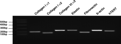
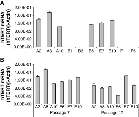
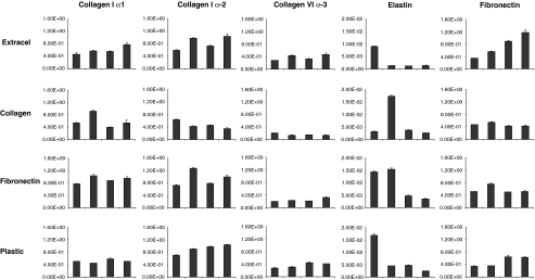
Tissue regeneration of the vocal fold lamina propria extracellular matrix (ECM) will be facilitated by the use of suitable vocal fold fibroblast (VFF) cell lines in appropriate model systems. Primary human VFFs (hVFFs) were steadily transduced by a retroviral vector containing human telomerase reverse transcriptase (hTERT) gene; immortalized cells grew and divided vigorously for more than 120 days. Biochemical characterization of the six transduced lines included, at different time points, expression of hTERT, telomerase activity, telomere lengths, and transcript levels of ECM constituents. Telomere lengths of the transfected lines were elongated and stable. Gene expression levels of collagen Ialpha1, collagen Ialpha2, collagen VIalpha3, elastin, and fibronectin were measured between the transduced cell clones and the primary hVFFs to verify transcription. Absence of inter- and intraspecies contamination was confirmed with DNA fingerprinting and karyotype analysis. Cell morphology, growth, and transcription expression were examined on 2D scaffolds-collagen, fibronectin, and hyaluronic acid. Immortalized hVFFs demonstrated normal attachment and spread on 2D scaffolds. Collagen Ialpha1, collagen Ialpha2, collagen VIalpha3, elastin, and fibronectin transcript expression was measured from immortalized hVFFs, for all surfaces. This is the first report of immortalization and biochemical characterization of hVFFs, providing a novel and invaluable tool for tissue regeneration applications in the larynx.


Figures

FIG. 1.
FIG. 1.
Electrophoresis of real-time PCR primers. PCR product size of each gene matched the predicted sizes as in Table 1.
FIG. 2.
FIG. 2.
hTERT gene expression levels of transduced hVFF lines by RT-PCR. hTERT mRNA data were normalized by housekeeping gene β-actin. (A) hTERT gene mRNA level of hTERT-transduced hVFF lines and control vector–transduced cell lines at passage 7. (B) hTERT gene mRNA of hTERT-transduced hVFF lines at passages 7 and 17.
FIG. 3.
FIG. 3.
Expression of exogenous telomerase activity in two independent parental normal hVFFs (21T and 59T hVFF) and their transduced cell lines. The treatment of the cells was described in Materials and Methods section. One of three independent TRAP assays is shown. (A) Quantification telomeric repeat amplification protocol assay of parental (21T and 59T), hTERT-transduced clones (A2, A8, A10, E6, E7, and E10), and vector-transduced (B1, B3, F1, and F5). Cellular extracts at passage 7 were tested for telomerase activity. Telomerase activity levels were normalized to the level of the internal control amplification products. (B) Telomeric repeat amplification protocol assay of hTERT-transduced hVFF cell clones at passages 7, 13, and 17.
FIG. 4.
FIG. 4.
Southern blot (telomere length assay) of transduced cell lines and parental hVFFs. Genomic DNA was isolated from hTERT-transduced populations, control, and uninfected parental lines at passages 8 and 18, and then hybridized with a telomeric probe to visualize the TRF. The first lane shows the molecular weight marker in kb. (A) At passage 8, telomere length of the cell lines established from 21T and 59T strains and telomeres of hTERT-positive clones (A2, A8, A10, E6, E7, and E10) were 19–20 kb, much longer than that hTERT-negative clones (B1, B3, F1, and F5) and parental cells (21T and 59T) whose range was 7.0–8.0 kb. The broad band of hTERT-negative and parental cells indicates wide variability of telomere length, while hTERT-transduced cells are more uniform. (B) Telomere length of hTERT-transduced cell lines and parental hVFFs at passages 8 and 18. Comparing the parental 21T and 59T, all of hTERT-positive cell lines maintained stable telomere length at 19–20 kb until passage 18.
FIG. 5.
FIG. 5.
Growth curves of transduced cell clones established from 21T hVFFs. (A) Growth curves of hTERT-positive cells (A2, A8, and A10) and control cells (B1 and B3). (B) PDs of hTERT-transduced single cell clones (A2, A8, and A10) and control vector–transduced cell lines (B1 and B3) are shown over 120 days of culture. X-axis indicates incubation days, and Y-axis indicates the number of population doublings.
FIG. 6.
FIG. 6.
Growth curves of transduced cell clones established from 59T hVFFs. (A) Growth curves of hTERT-positive cells (E6, E7, and E10) and control cells (F1 and F5). (B) PDs of hTERT-transduced single-cell clones (E6, E7, and E10) and control vector–transduced cell lines (F1 and F5) are shown over 120 days of culture. X-axis indicates incubation days, and Y-axis indicates the number of population doublings.
FIG. 7.
FIG. 7.
Morphologic appearance of parental hVFF cells, hTERT-transduced single clones (A2 and E6), and control empty vector–transduced single clones (B1 and F1) in growth phase.
FIG. 8.
FIG. 8.
Expression of collagen I α-1 (A), collagen I α-2 (B), collagen VI α-3 (C), fibronectin (D), and elastin (E) genes in hTERT-transduced cell clones (A2, A8, A10, E6, E7, and E10), control vector–transduced cell clones (B1, B3, F1, and F5), and parental hVFFs (21T and 59T). mRNA levels (ng/μL) were normalized by housekeeping gene β-actin.
FIG. 9.
FIG. 9.
Cytogenetic analysis of immortalized hVFF cell lines, showing all transduced fibroblast lines with normal human karyotypes. (A) Karyotype of a cell from 21T male donor (passage 5). (B) Karyotype of a cell from 59T female donor (passage 5).
FIG. 10.
FIG. 10.
Expression of collagen I α-1, collagen I α-2, collagen VI α-3, fibronectin, and elastin genes in hTERT immortalized cell clones, A8 hVFFs on Extracel, collagen, fibronectin, and plastic surfaces. mRNA levels (ng/μL) were normalized by housekeeping gene β-actin.

References

    1. Verdolini K. Ramig L. Review: occupational risks for voice problems. Logoped Phoniatr Vocol. 2001;26:37. - PubMed
    1. Ma E.P. Yiu E.M. Voice activity and participation profile: assessing the impact of voice disorders on daily activities. J Speech Lang Hear Res. 2001;44:511. - PubMed
    1. Benninger M.S. Alessi D. Archer S. Bastian R. Ford C. Koufman J. Sataloff R.T. Spiegel J.R. Woo P. Vocal fold scarring: current concepts and management. Otolaryngol Head Neck Surg. 1996;115:474. - PubMed
    1. Hirano M. Jackson C. Lecture. Phonosurgery: past, present, and future. American Broncho-Esophagological. 1995:Association.
    1. Johnson P.C. Mikos A.G. Fisher J.P. Jansen J.A. Strategic directions in tissue engineering. Tissue Eng. 2007;13:2827. - PubMed

Publication types

MeSH terms