

doi: 10.1016/j.tig.2008.11.006.
Epub 2008 Dec 26.
Evolutionary mutant models for human disease
Affiliations
- PMID: 19108930
- PMCID: PMC2828043
- DOI: 10.1016/j.tig.2008.11.006
Item in Clipboard
Evolutionary mutant models for human disease
Trends Genet.
2009 Feb.
Abstract
Although induced mutations in traditional laboratory animals have been valuable as models for human diseases, they have some important limitations. Here, we propose a complementary approach to discover genes and mechanisms that might contribute to human disorders: the analysis of evolutionary mutant models in which adaptive phenotypes mimic maladaptive human diseases. If the type and mode of action of mutations favored by natural selection in wild populations are similar to those that contribute to human diseases, then studies in evolutionary mutant models have the potential to identify novel genetic factors and gene-by-environment interactions that affect human health and underlie human disease.
Figures
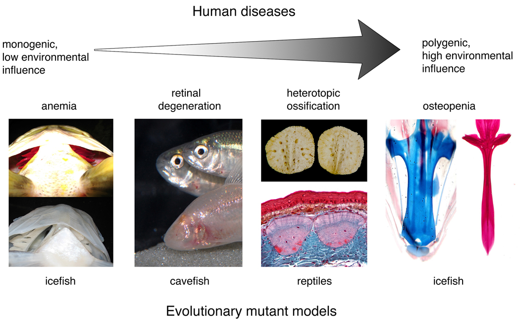
Evolutionary mutant models have the potential to reveal novel insight into the genetic basis of an array of different types of human diseases, from simple to complex. An example of a natural system that models a simple human disease is anemia in icefish. Note that the icefish (bottom panel) lacks red blood cells (as seen in the gills) compared to the closely related rockcod (top panel). Evolutionary mutants can also model complex human diseases including osteopenia in icefish. Note that the base of the icefish neurocranium is cartilaginous (blue, left panel) compared to the closely related rockcod, which has a mineralized neurocranium (red, right panel). Evolutionary mutants will also be useful for diseases in the middle of the continuum that are affected by alleles of major effect, but whose expression is complex and variable. Diseases of this type include retinal degeneration and heterotopic ossification, which are modeled by blind cavefish and reptile osteoderms, respectively. Cavefish image reproduced with permission from R. Borowsky. Osteoderm image courtesy of M. Vickaryous.
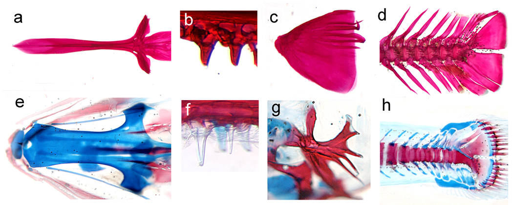
The evolutionary history of certain Antarctic notothenioid fish species has led to changes in the musculoskeletal system to increase buoyancy. Among other adaptations, several species have evolved bone loss, and now possess greatly reduced bony skeletons. These evolutionary adaptations model human diseases of decreased bone mineralization including osteopenia. An example of an osteopenia notothenioid species is Pseudochaenichthys georgianus (e–h), which, compared to the closely related but robustly mineralized species, Notothenia rossii (a–d), shows reduced levels of bone mineralization in the base of the skull (a,e), oral teeth (b,f), opercular bone (c,g), and caudal skeleton. Images show the staining of cartilage with alcian blue and bone with alizarin red in juvenile fish.
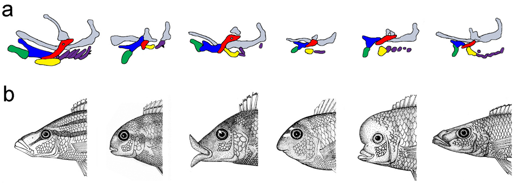
(a) Chemically induced zebrafish craniofacial mutants (wild-type configuration is to the left). Shaded shapes represent different pharyngeal cartilage elements depicted in the lateral view with the rostral-caudal axis running from left to right. Induced laboratory mutants have been useful for deducing the factors that pattern the embryonic craniofacial skeleton, including (left to right following the wild-type configuration) low/tfap2a, fla/pold1, bab/rerea, and her [26]. Note differences in the number, size, and orientation of elements. Gray elements depict the skull cartilages, green is the lower jaw, blue in the upper jaw, red in the hyosympletic cartilage, yellow is the ceratohyal cartilage, and purple elements are the posterior gill cartilages. Reproduced with permission of the Company of Biologists. (b) A series of evolutionary mutants illustrates the striking difference in craniofacial shape among closely related cichlid species. Here, natural selection has screened random mutations for differences in adult feeding morphology. These evolutionary mutants model both normal and clinical variation in human craniofacial architecture. Cichlid illustrations were drawn by R.C.A.
References
-
- Carroll SB, et al. From DNA to Diversity: Molecular Genetics and the Evolution of Animal Design, 2nd edition. Malden, MA, USA: Blackwell; 2005.
-
- Donovan A, et al. Positional cloning of zebrafish ferroportin1 identifies a conserved vertebrate iron exporter. Nature. 2000;403:776–781. - PubMed
-
- Lamason RL, et al. SLC24A5, a putative cation exchanger, affects pigmentation in zebrafish and humans. Science. 2005;310:1782–1786. - PubMed
-
- Sabherwal N, et al. Long-range conserved non-coding SHOX sequences regulate expression in developing chicken limb and are associated with short stature phenotypes in human patients. Hum. Mol. Genet. 2007;16:210–222. - PubMed
Publication types
MeSH terms
Grants and funding
LinkOut - more resources
Full Text Sources
