. 2009 Jan 1;182(1):489-97.
doi: 10.4049/jimmunol.182.1.489.

Toxoplasma gondii prevents chromatin remodeling initiated by TLR-triggered macrophage activation

Affiliations

Toxoplasma gondii prevents chromatin remodeling initiated by TLR-triggered macrophage activation

Jin Leng et al. J Immunol. .

Abstract

Macrophages infected with the opportunistic protozoan Toxoplasma gondii are unable to up-regulate many proinflammatory cytokine genes, including TNF (TNF-alpha), upon stimulation with LPS and other TLR ligands. In this study, we examined the influence of T. gondii on transcription factors associated with TNF-alpha transcription, as well as phosphorylation and acetylation of histone H3 at distal and proximal regions of the TNF-alpha promoter. During LPS stimulation, we found that Toxoplasma blocks nuclear accumulation of transcription factor c-Jun, but not that of cAMP response element-binding protein or NF-kappaB. However, chromatin immunoprecipitation studies revealed that binding of all of these transcription factors to the TNF promoter was decreased by T. gondii infection. Furthermore, the parasite blocked LPS-induced Ser(10) phosphorylation and Lys(9)/Lys(14) acetylation of histone H3 molecules associated with distal and proximal regions of the TNF-alpha promoter. Our results show that Toxoplasma inhibits TNF-alpha transcription by interfering with chromatin remodeling events required for transcriptional activation at the TNF promoter, revealing a new mechanism by which a eukaryotic pathogen incapacitates proinflammatory cytokine production during infection.


Conflict of interest statement

Disclosures

The authors have no financial conflict of interest.

Figures
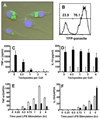
FIGURE 1
FIGURE 1. T. gondii inhibits LPS-induced TNF-α, but not IL-10 production
A and B, Bone marrow-derived macrophages were infected (4:1 ratio of tachyzoites:cells) with a T. gondii RH strain genetically engineered to express tandem copies of yellow fluorescence protein (YFP). After 12 h, cells were prepared for fluorescence microscopy (A) and flow cytometry (B). C and D, Twelve-hour-infected macrophages were stimulated with LPS (■) or cultured in medium alone (□), and then supernatants were collected 6 h later for TNF-α (C) and IL-10 (D) ELISA. E and F, Macrophages were infected (4:1 ratio of parasites: cells) and then 12 h later subjected to LPS stimulation. At the indicated time points, RNA was extracted, reverse transcribed into cDNA, and real-time PCR was performed for TNF-α (E) and IL-10 (F) transcripts. The data are normalized to GAPDH expression levels. E and F, ■ and □ indicate LPS-induced responses of infected and noninfected cells, respectively.
FIGURE 2
FIGURE 2. T. gondii inhibits TNF-α induction by multiple TLR ligands and multiple parasite strains inhibit TNF-α induction
A, Bone marrow-derived macrophages were infected with RH strain tachyzoites for 12 h, followed by a 6-h stimulation with LPS, Pam3Cys, or CpG. Supernatants were collected for subsequent TNF ELISA. B, Macrophages were infected with RH, CC, ENT, or DEG strain tachyzoites for 12 h, followed by a 6-h stimulation with LPS. Supernatants were collected for subsequent TNF ELISA.
FIGURE 3
FIGURE 3. T. gondii blocks RNA pol II recruitment to the TNF gene
Macrophages were infected with RH strain tachyzoites for 12 h, followed by a 30-min stimulation with LPS. Nuclear extracts were prepared and subjected to a ChIP assay using Ab against RNA pol II. A, ChIP DNA was amplified by standard PCR methodology using primers spanning the 5′ region of the TNF gene, and input DNA was amplified as a control. B and C, ChIP DNA was amplified by real-time PCR using two different primer sets spanning the 5′ end of the TNF gene. The data was normalized to input DNA and amplification was expressed as relative enrichment compared with cells in medium (defined as 1). Med, Cells incubated in medium alone; Tg, cells infected with Toxoplasma. *, p < 0.01 comparing LPS and Tg + LPS.
FIGURE 4
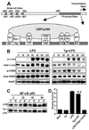
FIGURE 4. T. gondii blocks LPS-induced nuclear translocation of c-Jun, but not that of CREB or NF-κB p65
A, Schematic map of the mouse TNF promoter showing distal and proximal sites involved in transcriptional initiation. B, Macrophages were either infected or left uninfected for 12 h and then subjected to LPS stimulation for the indicated time periods (min). Western blotting for total and activated forms of c-Jun and CREB was performed on nuclear lysates. Blotting for the nuclear enzyme poly(ADP-ribose) polymerase was performed to confirm equal protein loading. C, Cytoplasmic and nuclear extracts were prepared from infected and noninfected macrophages stimulated with LPS for the indicated time periods (min). The extracts were subsequently probed with Ab to NF-κB p65. In D, the in vivo-binding capability of nuclear NF-κB p65 was determined using an ELISA-based method to measure transcription factor binding to solid-phase target oligonucleotides. In this panel, addition of soluble target oligonucleotide (Oligo Comp) blocked p65 binding, confirming the specificity of the assay. Med, Cells cultured in medium alone; Tg, T. gondii-infected cells; n.s., not significant comparing LPS and Tg plus LPS.
FIGURE 5
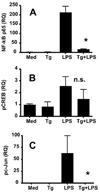
FIGURE 5. T. gondii blocks LPS-induced binding of phosphorylated c-Jun and NF-κB p65 to the TNF promoter
Bone marrow-derived macrophages were infected with RH strain tachyzoites and then 12 h later subjected to a 30-min LPS stimulation. ChIP assays were performed using Abs specific for NF-κB p65 (A), phospho-CREB (B), and phospho-c-Jun (C). The immunoprecipitated DNA was amplified by real-time PCR using primers spanning either proximal (for c-Jun and CREB) or distal (for NF-κB p65) sites of the TNF promoter. The results were normalized to the input DNA and expressed as relative quantitation (RQ) where medium is defined as 1. Med, Medium; Tg, cells infected with T. gondii; n.s., not significant. *, p < 0.01 comparing LPS and Tg plus LPS.
FIGURE 6
FIGURE 6. T. gondii globally blocks LPS-induced phosphorylation of histone H3 at Ser10 but has no effect on Lys9/14 histone H3 acetylation
A, Macrophages were infected and then 12 h later subjected to LPS stimulation for the indicated time periods (min). Total cell lysates were extracted and subjected to Western blotting using Abs specific to phospho-Ser10 histone H3, acetyl Lys9/14, and total H3. In B, fluorescence microscopy was used to examine Lys9/14 acetylation (top panels) and Ser10 phosphorylation (bottom panels) of histone H3 in infected and noninfected cells stimulated for 30 min with LPS. Macrophages were stained with Ab specific for acetylated or phosphorylated histone H3 as indicated (red) and Toxoplasma p30 (green). Nuclei are stained with 4′,6-diamidino-2-phenylindole (blue). In C, cells that were positive for phospho-Ser10 at histone H3 staining were counted in each group of cells (~200 cell/condition). *, p < 0.01 comparing infected and noninfected cells. D, Twelve-hour-infected and control macrophages were stimulated with LPS (100 ng/ml) and cell lysates were prepared at the indicated time points (min). Immunoblotting for phospho-MSK1 was subsequently performed. As a control for protein loading, levels of histone H3 were assessed in the same samples. Med, Medium; Tg, T. gondii.
FIGURE 7
FIGURE 7. T. gondii blocks LPS-induced histone H3 modification at the TNF promoter
Macrophages were infected with T. gondii and then 12 h later subjected to LPS stimulation for 30 min. The cells were fixed with formaldehyde and a ChIP assay was performed using Abs specific for Ser10 phosphorylated (A and C) or Lys9/14 acetylated (B and D) histone H3. The resulting DNA was amplified by real-time PCR using primers spanning either proximal (A and B) or distal (C and D) sites of the TNF promoter region. As a control, the same ChIP DNA was amplified using primers spanning the IL-10 promoter region (E and F). The results were normalized to the input DNA and expressed as relative quantitation (RQ), with medium defined as 1. Med, Cells incubated in medium alone; Tg, cells infected with Toxoplasma. *, p < 0.05 comparing LPS and Tg plus LPS.
FIGURE 8
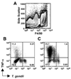
FIGURE 8. T. gondii inhibits TNF-α production during in vivo infection of F4/80-positive macrophages
Mice were infected by i.p. injection of 106 RH strain tachyzoites. After 12 h, cells in the peritoneal cavity were collected and cultured for 6 h in either medium alone or LPS in the presence of brefeldin A. Cells were then subjected to surface staining for F4/80 and intracellular staining for TNF-α and Toxoplasma p30 (SAG-1) to detect tachyzoites. A, F4/80-positive cells in the peritoneal cavity. The rectangle delineates cells selected for analysis. B, TNF-α and infection levels in F4/80-positive cells cultured in medium. C, TNF-α and infection levels in F4/80-positive cells stimulated in vitro with LPS.

References

    1. Alexander J, Hunter CA. Immunoregulation during toxoplasmosis. Chem. Immunol. 1998;70:81–102. - PubMed
    1. Denkers EY, Gazzinelli RT. Regulation and function of T cell-mediated immunity during Toxoplasma gondii infection. Clin. Microbiol. Rev. 1998;11:569–588. - PMC - PubMed
    1. Gazzinelli RT, Wysocka M, Hieny S, Scharton-Kersten T, Cheever A, Kuhn R, Muller W, Trinchieri G, Sher A. In the absence of endogenous IL-10, mice acutely infected with Toxoplasma gondii succumb to a lethal immune response dependent upon CD4+ T cells and accompanied by overproduction of IL-12, IFN-γ, and TNF-α. J. Immunol. 1996;157:798–805. - PubMed
    1. Butcher BA, Kim L, Johnson PF, Denkers EY. Toxoplasma gondii tachyzoites inhibit proinflammatory cytokine induction in infected macrophages by preventing nuclear translocation of the transcription factor NF-κB. J. Immunol. 2001;167:2193–2201. - PubMed
    1. Lee CW, Bennouna S, Denkers EY. Screening for Toxoplasma gondii regulated transcriptional responses in LPS-activated macrophages. Infect. Immun. 2006;74:1916–1923. - PMC - PubMed

Publication types

MeSH terms

LinkOut - more resources