. 2009 Jan 1;182(1):604-12.
doi: 10.4049/jimmunol.182.1.604.

Crosstalk between CXCR4/stromal derived factor-1 and VLA-4/VCAM-1 pathways regulates neutrophil retention in the bone marrow

Affiliations

Crosstalk between CXCR4/stromal derived factor-1 and VLA-4/VCAM-1 pathways regulates neutrophil retention in the bone marrow

Joseph M Petty et al. J Immunol. .

Abstract

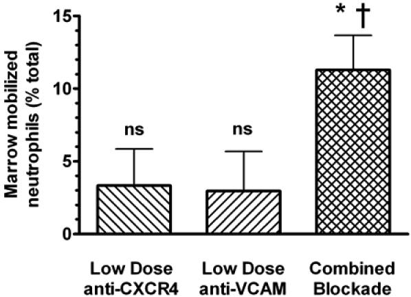
Neutrophil retention in and release from the bone marrow is a critical process that remains incompletely understood. Previous work has implicated the CXCR4/stromal derived factor-1 (SDF-1) chemokine axis in the marrow retention of neutrophils, yet the adhesion pathways responsible for this retention are unknown. Because alpha(4)beta(1) integrin (VLA-4) and its ligand VCAM-1 play a central role in the interactions of hematopoietic stem cells, lymphocytes, and developing neutrophils in the marrow, we investigated whether this integrin might be involved in marrow neutrophil retention and release. In this study, we show that VLA-4 is expressed on murine marrow neutrophils and decreases with maturation, whereas blockade of this integrin leads to the release of marrow neutrophils. Marrow neutrophils adhere via VLA-4 to VCAM-1, which is expressed on marrow endothelium and stroma, and inhibition of VCAM-1 causes release of marrow neutrophils. Furthermore, SDF-1 (CXCL12) signaling through neutrophil CXCR4 augments VLA-4 adhesion to VCAM-1 in vitro, an effect that is blocked by preincubation with pertussis toxin. In vivo blockade of both CXCR4 and alpha(4) causes synergistic release of marrow neutrophils, showing that cross-talk between CXCR4 and VLA-4 modulates marrow retention of these cells. Taken together, these results indicate that the VLA-4/VCAM adhesion pathway is critical in the retention and maturation-controlled release of neutrophils from the marrow, while providing an important link between the CXCR4/SDF-1 signaling axis and the adhesion events that govern this process.


Figures

Figure 1
Figure 1. Murine neutrophil expression of surface α4 integrin in the context of α4β1 (VLA-4) decreases with maturation
(A) Murine bone marrow and blood leukocytes were stained with Abs against α4 (CD49d) and Gr-1 (a murine marker of neutrophils) and analyzed by flow cytometry, gating for Gr-1 positive cells. (B) As α4 is expressed as one of two heterodimers (α4β1 and α4β7), murine marrow neutrophils were also stained with Ab specific for LPAM (the α4β7 heterodimer) to rule out significant LPAM expression. (C) Relative α4 expression on marrow and blood neutrophils was compared using relative fluorescence index (RFI) expressed as a ratio of the mean fluorescence intensity of cells stained with α4 Ab versus isotype control Ab. (D) Correlation between marrow neutrophil maturation state and expression of α4 was examined using in vivo pulse labeling with the thymidine analog BrdU (see Methods). In this technique cells that are post-mitotic at the time of pulse (the most mature cells) do not incorporate BrdU (BrdUnegative), whereas less mature, dividing cells incorporate BrdU. These cells are then detected as BrdUbright (more mature) or BrdUdim (least mature) depending on the number of subsequent cell divisions prior to entry into the post-mitotic marrow neutrophil pool. Marrow cells were analyzed 24h after BrdU pulse, and flow cytometry was gated for Gr-1 expression and BrdU staining intensity. α4 staining in the cell populations is expressed as relative fluorescence index to isotype control. Data points from all experiments are the means of 3-5 separate experiments performed for each analyzed population of cells (± SEM). Expression significantly different when compared to each other by *t-test (p<0.001) or **one-way ANOVA (p=0.001).
Figure 2
Figure 2. VLA-4 integrin blockade results in significant neutrophil release from bone marrow
(A) Mice were infused with either CD49d (α4) neutralizing or isotype control Abs (30μg) and blood neutrophil levels were measured 4h later. (B) This effect was further examined using neutrophil adoptive transfer in which 111indium-labeled marrow neutrophils were injected into naïve mice and allowed to localize to marrow for 4h before infusion of anti-CD49d or control Ab. Thirty minutes after Ab infusion, blood and tissue 111In-neutrophil content was determined by gamma counting. (C) The role of α4 in neutrophil homing to marrow was investigated in mice injected with blocking Ab or isotype control 30 min prior to labeled cell infusion. Blood and tissue 111In-neutrophil content was determined 4h after cell infusion. (D) To determine whether the effects of α4 blockade reflect disruption of α4β1 (VLA-4) or α4β7 (LPAM), neutralizing Ab specific for LPAM (30μg) was used in similar homing experiments. (E) To examine the nonspecific effects of antibody binding of neutrophil cell surface antigens, blocking antibodies against CD62L (L-selectin) or CD11a (αL) (30 ug) were infused in separate neutrophil adoptive transfer experiments (similar to those reported in panel B) and compared to isotype control Ab infusions. Marrow levels of labeled neutrophils are shown. Data points are the means of 3 to 5 mice/condition (± SEM). Significantly different when compared to control-treated animals, * p<0.005, **p<0.02; ns indicates not significant. (F) To determine whether neutrophil might be activated by binding of the CD49d blocking Ab, calcium flux assays were performed. Cells were labeled with the fluorochrome Indo-1/AM and calcium mobilization in response to α4-blocking antibody (20 ug/ml) or fMLP (1μM; as a positive control) was determined using flow cytometry.
Figure 3
Figure 3. Marrow neutrophil sensitivity to VLA-4 neutralization increases with cell maturation
Endogenous marrow neutrophil mobilization was examined using in vivo pulse labeling with the thymidine analog BrdU (see Methods) 48h prior to injection of either CD49d neutralizing or isotype control Abs. Mice were then euthanized 4h after Ab injection and blood and marrow analyzed by hematology analyzer and flow cytometry gating for either Gr-1hi (blood) or Gr-1+ (marrow) cells (neutrophils), as well as BrdU staining. In this technique three populations of neutrophils are identified: BrdUnegative (most mature), BrdUbright (less mature), and BrdUdim (least mature) (see Fig Legend 1). (A) Marrow neutrophil levels in anti-CD49d and control Ab-treated animals. Data is expressed as percentage of total Gr-1+ cells, and is normalized for total differences in total marrow cells (per femur). (B) Blood neutrophil levels. Data is expressed as absolute blood levels for each population. Data points are the means of 3 to 5 mice/condition (± SEM). *Significantly different when compared to control-treated animals, p<0.05; ns indicates not significant.
Figure 4
Figure 4. Marrow neutrophils adhere to VCAM-1 through VLA-4
(A) Isolated murine marrow neutrophils were labeled with 111In and incubated for 30min at 37°C in tissue culture wells coated with varying concentrations of VCAM-1, after which the wells were washed, and the cell content of each well was assayed by gamma counting and expressed as a percentage of the total amount plated. (B) Labeled neutrophils were preincubated with α4 or α4β7 (LPAM) neutralizing Abs (15 μg/ml) for 30min prior to VCAM-1 adhesion and analysis. Data points are the means of 3 to 5 separate experiments (± SEM). *Significantly different when compared to isotype control, p<0.0001; ns indicates not significant.
Figure 5
Figure 5. VCAM-1 is expressed on stromal and endothelial cells in murine bone marrow
Fluorescent immunohistology for VCAM-1 was performed on fixed whole bone marrow plugs from mouse femurs. (A) VCAM-1 expression (green) appears to localize to the venous endothelium (white arrow) and large interdigitating stromal cells (yellow arrow). Blue staining (DAPI) indicates cell nuclei. (B) Secondary-control staining. Magnification 400x.
Figure 6
Figure 6. VCAM-1 blockade releases neutrophils from bone marrow
(A) Mice were infused with either VCAM-1 (CD106) neutralizing or isotype control Abs (30μg) and blood neutrophil levels were measured 4h later. (B) This effect was further examined using adoptive transfer of 111In-labeled neutrophils 4h before infusion of anti-VCAM-1 or control Ab. Thirty minutes after Ab infusion, blood and tissue 111In-neutrophil content was determined by gamma counting. (C) The role of VCAM-1 in neutrophil homing to marrow was investigated using mice injected with blocking Ab or isotype control Ab 30 min prior to labeled cell infusion. Blood and tissue 111In-neutrophil content was determined 4h after cell infusion. (D) To examine the nonspecific effects of antibody binding of marrow cell surface antigens in this model, blocking antibody against CD54 (ICAM-1) (30 ug) was infused in a separate neutrophil adoptive transfer experiment (similar to those reported in panel B) and compared to isotype control Ab infusion. Marrow levels of labeled neutrophils are shown. Data points are the means of 3 to 5 mice/condition (± SEM). Significantly different when compared to control-treated animals, * p<0.005; ns indicates not significant.
Figure 7
Figure 7. Neutrophil VLA-4/VCAM-1 adhesion is modulated by SDF-1/CXCR4 signaling in vitro
(A) Neutrophil adhesion to VCAM-1 coated wells was performed as described in the presence or absence of co-immobilized SDF-1. (B) Labeled neutrophils were incubated overnight with the Gi protein-coupled signaling inhibitor pertussis toxin (PTX) prior to VCAM adhesion. Data points are the means of 4-5 separate experiments (± SEM). * Significantly different, p<0.0001; ns indicates not significant.
Figure 8
Figure 8. SDF-1 is closely co-localized with VCAM-1 on the surface of bone marrow stromal cells
Dual fluorescent immunohistology for SDF-1 and VCAM-1 was performed on fixed whole bone marrow plugs from mouse femurs. Samples were imaged by fluorescent microscopy for each fluorophore individually and together. (A) SDF-1. Red fluorescence denotes SDF-1 staining (white arrowheads indicate cells staining solely for SDF-1). (B) VCAM-1. Green fluorescence indicates VCAM-1 staining (while light blue arrowheads indicate cells staining solely for VCAM-1). (C) Merged image. Yellow fluorescence indicates large areas of co-localization between SDF-1 and VCAM; yellow arrowheads indicate examples of co-localization. (D) Dual secondary-control staining. Blue staining (DAPI) indicates cell nuclei. Magnification 400x.
Figure 9
Figure 9. Neutralization of CXCR4 and VCAM-1 is synergistic in vivo
The apparent crosstalk between CXCR4 and VLA-4 was examined in vivo. Mice were treated with low doses of either VCAM blocking or CXCR4 blocking Abs, both Abs simultaneously, or isotype control 4h after labeled marrow neutrophils were sequestered to the marrow, as described. Subsequent marrow content of labeled neutrophils was assayed 2h after antibody infusion. Results are expressed as percent decrease compared to control-treated animals. Data points are the means of 5 separate experiments (± SEM). *Significantly different when compared to control treated animals, p<0.01; †Significantly different when compared to low dose VCAM blocking Ab or low dose CXCR4 blocking Ab alone, p<0.05; ns, not significantly different compared to control treated animals.

References

    1. Opdenakker G, Fibbe WE, Van Damme J. The molecular basis of leukocytosis. Immunol Today. 1998;19:182–189. - PubMed
    1. Suratt BT, Petty JM, Young SK, Malcolm KC, Lieber JG, Nick JA, Gonzalo JA, Henson PM, Worthen GS. Role of the CXCR4/SDF-1 chemokine axis in circulating neutrophil homeostasis. Blood. 2004;104:565–571. - PubMed
    1. Ma Q, Jones D, Springer TA. The chemokine receptor CXCR4 is required for the retention of B lineage and granulocytic precursors within the bone marrow microenvironment. Immunity. 1999;10:463–471. - PubMed
    1. Martin C, Burdon PC, Bridger G, Gutierrez-Ramos JC, Williams TJ, Rankin SM. Chemokines acting via CXCR2 and CXCR4 control the release of neutrophils from the bone marrow and their return following senescence. Immunity. 2003;19:583–593. - PubMed
    1. Teixido J, Hemler ME, Greenberger JS, Anklesaria P. Role of beta 1 and beta 2 integrins in the adhesion of human CD34hi stem cells to bone marrow stroma. J Clin Invest. 1992;90:358–367. - PMC - PubMed

Publication types

MeSH terms