. 2009 Jan 1;182(1):623-35.
doi: 10.4049/jimmunol.182.1.623.

CD11b+ myeloid cells are the key mediators of Th2 cell homing into the airway in allergic inflammation

Affiliations

CD11b+ myeloid cells are the key mediators of Th2 cell homing into the airway in allergic inflammation

Benjamin D Medoff et al. J Immunol. .

Abstract

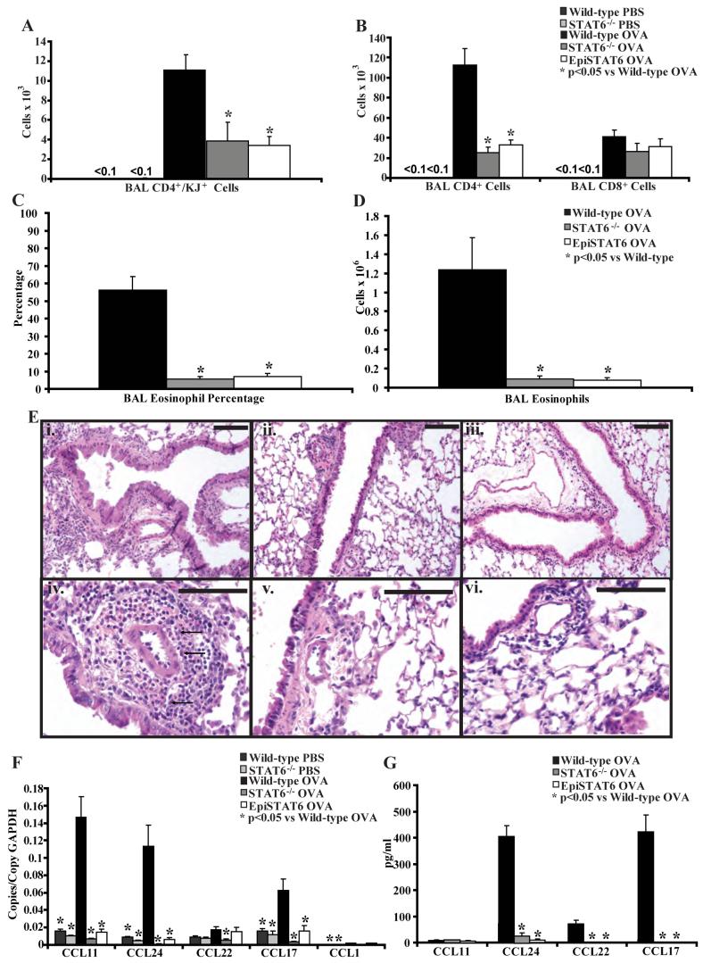
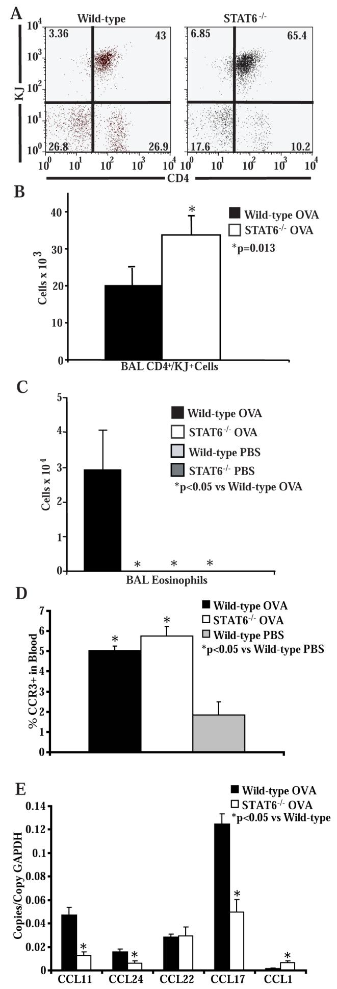
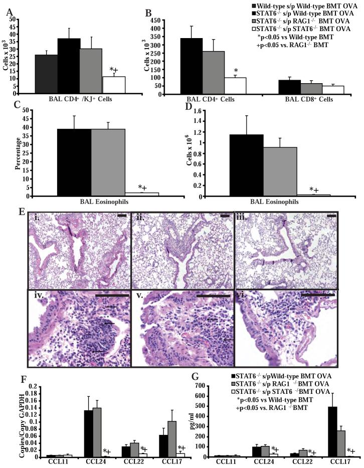
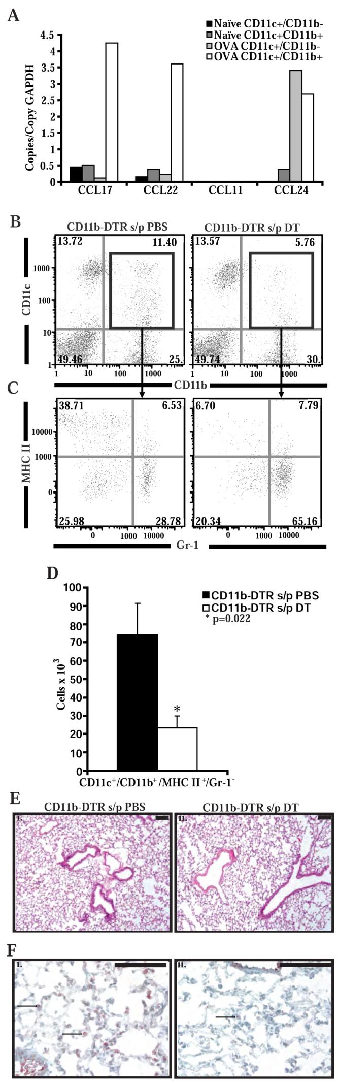
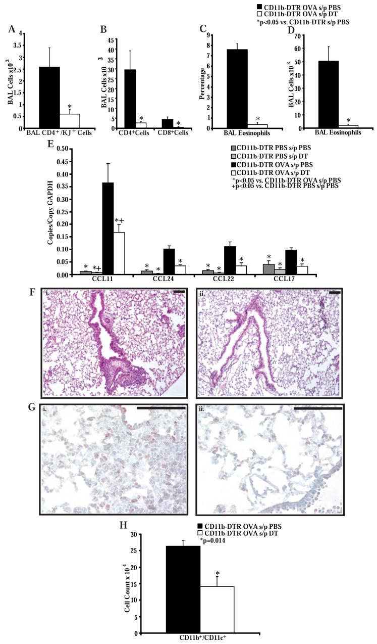
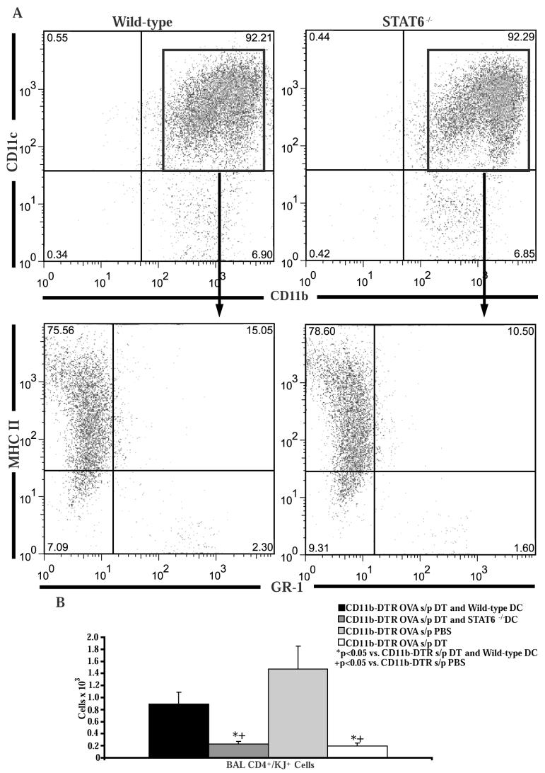
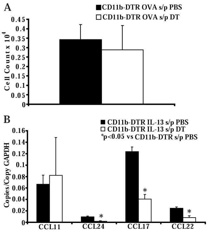
STAT6-mediated chemokine production in the lung is required for Th2 lymphocyte and eosinophil homing into the airways in allergic pulmonary inflammation, and thus is a potential therapeutic target in asthma. However, the critical cellular source of STAT6-mediated chemokine production has not been defined. In this study, we demonstrate that STAT6 in bone marrow-derived myeloid cells was sufficient for the production of CCL17, CCL22, CCL11, and CCL24 and for Th2 lymphocyte and eosinophil recruitment into the allergic airway. In contrast, STAT6 in airway-lining cells did not mediate chemokine production or support cellular recruitment. Selective depletion of CD11b(+) myeloid cells in the lung identified these cells as the critical cellular source for the chemokines CCL17 and CCL22. These data reveal that CD11b(+) myeloid cells in the lung help orchestrate the adaptive immune response in asthma, in part, through the production of STAT6-inducible chemokines and the recruitment of Th2 lymphocytes into the airway.


Figures

Figure 1
Figure 1. STAT6 expression in airway lining cells does not support Th2 cell and eosinophil trafficking into the airways
A) Number of CD4+ and KJ+ OVA-specific Th2 cells, and B) CD4+ and CD8+ T cells in the BAL of wild-type, STAT6-/- and EpiSTAT6 mice following adoptive transfer of OVA-specific in vitro polarized Th2 lymphocytes and 4 OVA or PBS challenges (n = 4-6 mice per group, from 2 experiments). C) Percentage and D) number of eosinophils in the BAL of wild-type, STAT6-/- and EpiSTAT6 mice following adoptive transfer of OVA-specific in vitro polarized Th2 lymphocytes and 4 OVA challenges (n = 6 mice per group, from 2 experiments). E) Representative lung histology (stained with hematoxylin and eosin) from wild-type (i, iv - 100x and 400x respectively), STAT6-/- mice (ii, v), and EpiSTAT6 (iii, vi) mice following adoptive transfer of OVA-specific in vitro polarized Th2 lymphocytes and 4 OVA challenges (n = 6 mice per group, from 2 experiments). Black bars are 100 μm, arrows demonstrate clusters of eosinophils only in the section from wild-type lungs. F) Lung chemokine RNA copies normalized to copies of GAPDH RNA and G) BAL chemokine protein levels in wild-type, STAT6-/- and EpiSTAT6 mice following adoptive transfer of OVA-specific in vitro polarized Th2 lymphocytes and 4 OVA or PBS challenges (n = 4-6 mice per group, from 2 experiments).
Figure 2
Figure 2. Mucus hyper-secretion in wild-type mice, STAT6-/- mice, EpiSTAT6 mice, and STAT6-/- mice following bone marrow transplant
Representative histologic sections stained with PAS to evaluate mucus hypersecretion following adoptive transfer of OVA-specific in vitro polarized Th2 lymphocytes and 4 OVA challenges. Sections are from wild-type BALB/c mice (A), STAT6-/- mice (B), EpiSTAT6 mice (C), wild-type BALB/c mice after bone marrow transplant with wild-type bone marrow (D), STAT6-/- mice after bone marrow transplant with wild-type bone marrow (E), or STAT6-/- mice after bone marrow transplant with RAG1-/- bone marrow (F). Black bars are 100 μm.
Figure 3
Figure 3. Intratracheal transfer of Th2 cells does not restore eosinophil trafficking into the airways in STAT6-/- mice
A) Representative flow cytometry of BAL cells stained for CD4 and KJ following intratracheal transfer of OVA-specific in vitro polarized Th2 lymphocytes and 3 OVA challenges (n = 6 mice per group, from 2 experiments). B) Number of CD4+ and KJ+ OVA-specific Th2 cells in the BAL of wild-type and STAT6-/- mice following intratracheal transfer of OVA-specific in vitro polarized Th2 lymphocytes and 3 OVA challenges (n = 6 mice per group, from 2 experiments). C) Number of eosinophils in the BAL of wild-type and STAT6-/-mice following intratracheal transfer of OVA-specific in vitro polarized Th2 lymphocytes and 3 OVA challenges (n = 6 mice per group, from 2 experiments). D) Percentage of CCR3+ cells in the granulocyte gate of cells isolated from blood of wild-type and STAT6-/- mice following intratracheal transfer of OVA-specific in vitro polarized Th2 lymphocytes and 3 OVA or PBS challenges (n = 3 mice per group, from 1 experiment). E) Lung chemokine RNA copies normalized to copies of GAPDH RNA in wild-type and STAT6-/- mice following intratracheal transfer of OVA-specific in vitro polarized Th2 lymphocytes and 3 OVA challenges (n = 6 mice per group, from 2 experiments).
Figure 4
Figure 4. STAT6 expression in myeloid derived cells in the lung does support Th2 cell and eosinophil trafficking into the airways
A) Number of OVA-specific Th2 cells in the BAL of wild-type BALB/c mice after bone marrow transplant with wild-type bone marrow or STAT6-/- mice after bone marrow transplant with wild-type, STAT6-/- or RAG1-/- bone marrow, and following adoptive transfer of OVA-specific in vitro polarized Th2 lymphocytes and 4 OVA challenges (n = 4-8 mice per group, from 3 experiments). B) Number of CD4+ T cells, and CD8+ T cells in the BAL of STAT6-/- mice after bone marrow transplant with wild-type, STAT6-/-, or RAG1-/- bone marrow, and following adoptive transfer of OVA-specific in vitro polarized Th2 lymphocytes and 4 OVA challenges (n = 8 mice per group, from 2 experiments). C) Percentage and D) number of eosinophils in the BAL of STAT6-/- mice after bone marrow transplant with wild-type, STAT6-/- or RAG1-/- bone marrow, and following adoptive transfer of OVA-specific in vitro polarized Th2 lymphocytes and 4 OVA challenges (n = 8 mice per group, from 2 experiments). E) Representative lung histology from STAT6-/- mice after bone marrow transplant with wild-type (i, iv - 100x and 400x respectively), STAT6-/- (ii, v), or RAG1-/- (iii, vi) bone marrow, and following adoptive transfer of OVA-specific in vitro polarized Th2 lymphocytes and 4 OVA challenges (n = 8 mice per group, from 2 experiments). Black bars are 100 μm. F) Lung chemokine RNA copies normalized to copies of GAPDH RNA and G) BAL chemokine protein levels in STAT6-/- mice after bone marrow transplant with wild-type, STAT6-/- or RAG1-/- bone marrow, and following transfer of OVA-specific in vitro polarized Th2 lymphocytes and 4 OVA challenges (n = 8 mice per group, from 2 experiments).
Figure 5
Figure 5. DT administration selectively depletes myeloid dendritic cells in CD11b-DTR mice
A) Chemokine RNA copies normalized to copies of GAPDH from CD11c+/CD11b+ and CD11c+/CD11b- cells sorted from the lungs of wild-type naïve mice and wild-type mice following adoptive transfer of OVA-specific in vitro polarized Th2 lymphocytes and 4 OVA challenges (n = 4 pooled mice per group, from 1 experiment). B) Representative flow cytometry of lung cells isolated from CD11b-DTR mice 4 days after 2 treatments with PBS or DT. Cells are stained with labeled antibodies to CD11b and CD11c and gated on the monocytes/macrophages. C) Representative flow cytometry of lung cells isolated from CD11b-DTR mice 4 days after 2 treatments with PBS or DT. Cells were stained with labeled antibodies to Gr-1 (RB6-8C5 from BD Pharmingen), MHC II, CD11b and CD11c and gated on monocytes/macrophages and on CD11b+/CD11c+ cells. D) Number of CD11c+/CD11b+/MHC-II+/Gr-1- cells in the lungs of CD11b-DTR mice as measured by flow cytometry after treatment with PBS or DT (n = 6 mice per group, from 2 experiments). E) Representative lung histology from CD11b-DTR mice 4 days after 2 treatments with PBS (i) or DT (ii). Lung sections are stained with hematoxylin and eosin. Black bar is 100 μm. F) Representative immunohistochemistry for GFP in lungs taken from CD11b-DTR mice 4 days after 2 treatments with PBS (i) or DT (ii).
Figure 6
Figure 6. DT administration to CD11b-DTR mice impairs Th2 cell and eosinophil recruitment into the airways in an adoptive transfer model of allergic airway inflammation
A) Number of OVA-specific Th2 cells and B) CD4+ and CD8+ T cells in the BAL of CD11b-DTR mice after treatment with PBS or DT and following adoptive transfer of OVA-specific in vitro polarized Th2 lymphocytes and 4 OVA challenges (n = 8 mice per group, from 3 experiments). C) Percentage and D) number of eosinophils in the BAL of CD11b-DTR mice after treatment with PBS or DT and following adoptive transfer of OVA-specific in vitro polarized Th2 lymphocytes and 4 OVA challenges (n = 8 mice per group, from 3 experiments). E) Lung chemokine RNA copies normalized to copies of GAPDH RNA in CD11b-DTR mice after treatment with PBS or DT and following adoptive transfer of OVA-specific in vitro polarized Th2 lymphocytes and 4 OVA or PBS challenges (n = 3-6 mice per group, from 2 experiments). F) Representative lung histology from CD11b-DTR mice treated with PBS (i) or DT (ii), and following adoptive transfer of OVA-specific in vitro polarized Th2 lymphocytes and 4 OVA challenges (n = 8 mice per group, from 3 experiments). Black bars are 100 μm. G) Representative immunohistochemistry for GFP in lungs taken from CD11b-DTR mice after treatment with PBS or DT and following adoptive transfer of OVA-specific in vitro polarized Th2 lymphocytes and 4 OVA challenges (n = 4 mice per group, from 1 experiment). Black bars are 10 μm. H) Number of CD11c+/CD11b+ cells in the lungs of CD11b-DTR mice as measured by flow cytometry after treatment with PBS or DT and following adoptive transfer of OVA-specific in vitro polarized Th2 lymphocytes and 4 OVA challenges (n = 8 mice per group, from 3 experiments).
Figure 7
Figure 7. Intratracheal transfer of bone marrow-derived dendritic cells restores Th2 cell recruitment in CD11b+ myeloid cell depleted CD11b-DTR mice
A) Bone marrow cells from wild-type or STAT6-/- mice were cultured for 10 days in media supplemented with GM-CSF. The non-adherent cells were harvested and stained with antibodies to CD11b, CD11c, Gr-1, and MHC II. B) Number of OVA-specific Th2 cells in the BAL following adoptive transfer of OVA-specific in vitro polarized Th2 lymphocytes and 4 OVA challenges in CD11b-DTR mice which had been injected with DT and had received intratracheal transfer of wild-type or STAT6-/- bone marrow derived dendritic cells (n = 10 mice per group, from 2 experiments). CD11b-DTR mice treated with DT or PBS following adoptive transfer of OVA-specific in vitro polarized Th2 lymphocytes and 4 OVA challenges (n = 8 mice per group, from 2 experiments) served as controls.
Figure 8
Figure 8. DT administration to CD11b-DTR mice does not impair Th2 cell accumulation in the lymph node but does reduce IL-13 induced chemokine production
A) Numbers of OVA-specific Th2 cells in the thoracic lymph nodes of CD11b-DTR mice after treatment with PBS or DT and following adoptive transfer of OVA-specific in vitro polarized Th2 lymphocytes and 4 OVA challenges (n = 5 mice per group, from 2 experiments). B) Lung chemokine RNA copies normalized to copies of GAPDH RNA in CD11b-DTR mice after treatment with PBS or DT and 24 hours following intranasal IL-13 administration (n = 6 mice per group, from 2 experiments).

References

    1. Busse WW, Lemanske RF., Jr. Asthma. N Engl J Med. 2001;344:350–362. - PubMed
    1. Larche M, Robinson DS, Kay AB. The role of T lymphocytes in the pathogenesis of asthma. J Allergy Clin Immunol. 2003;111:450–463. quiz 464. - PubMed
    1. Woodfolk JA. T-cell responses to allergens. J Allergy Clin Immunol. 2007;119:280–294. quiz 295-286. - PubMed
    1. Robinson DS, Hamid Q, Ying S, Tsicopoulos A, Barkans J, Bentley AM, Corrigan C, Durham SR, Kay AB. Predominant TH2-like bronchoalveolar T-lymphocyte population in atopic asthma. N Engl J Med. 1992;326:298–304. - PubMed
    1. Del Prete GF, De Carli M, D’Elios MM, Maestrelli P, Ricci M, Fabbri L, Romagnani S. Allergen exposure induces the activation of allergen-specific Th2 cells in the airway mucosa of patients with allergic respiratory disorders. Eur J Immunol. 1993;23:1445–1449. - PubMed

Publication types

MeSH terms