. 2009 Mar;83(6):2417-28.
doi: 10.1128/JVI.02392-08. Epub 2008 Dec 24.

Latent species C adenoviruses in human tonsil tissues

Affiliations

Latent species C adenoviruses in human tonsil tissues

C T Garnett et al. J Virol. 2009 Mar.

Abstract

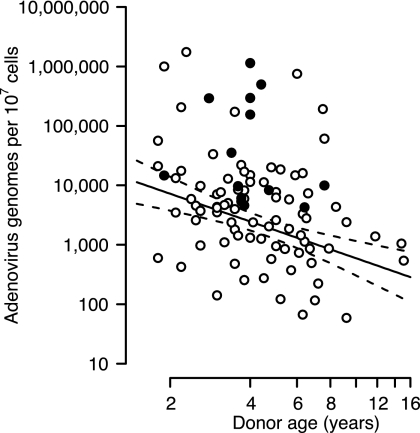
Although species C human adenoviruses establish persistent infections, the molecular details of this lifestyle remain poorly understood. We previously reported that adenovirus DNA is found in human mucosal T lymphocytes in a noninfectious form (C. T. Garnett, D. Erdman, W. Xu, and L. R. Gooding, J. Virol. 76:10608-10616, 2002). In this study, human tonsil and adenoid tissues were analyzed to determine the dynamics of infection, the rate of clearance of viral DNA, and the possibility of reactivation of virus from these tissues. The presence of viral DNA peaked at 4 years of age and declined thereafter. The average number of viral genomes declined with the age of the donor. The frequency of virus-bearing cells ranged from 3 x 10(-7) to 3.4 x 10(-4), while the amount of viral DNA per cell varied less, with an average of 280 copies per cell. All species C serotypes were represented in these tissues, although adenovirus type 6 was notably rare. Infectious virus was detected infrequently (13 of 94 of donors tested), even among donors with the highest levels of adenoviral DNA. Adenovirus transcripts were rarely detected in uncultured lymphocytes (2 of 12 donors) but appeared following stimulation and culture (11 of 13 donors). Viral DNA replication could be stimulated in most donor samples by lymphocyte stimulation in culture. New infectious virus was detected in 13 of 15 donors following in vitro stimulation. These data suggest that species C adenoviruses can establish latent infections in mucosal lymphocytes and that stimulation of these cells can cause viral reactivation resulting in RNA transcription, DNA replication, and infectious virus production.


Figures

FIG. 1.
FIG. 1.
The fraction of children with adenovirus DNA in lymphoid cells of the adenoids and tonsils peaks at approximately 4 years of age. Adenoid and tonsil samples from 202 donors sorted by age were partitioned into 18 age groups. The percentage of adenovirus DNA-positive samples in each group is plotted as a function of the median age for the group. The solid line is a best-fit second-order local polynomial regression curve.
FIG. 2.
FIG. 2.
The number of adenoviral genomes per lymphoid cell of the adenoids and tonsils declines with the age of the donor. The viral genome copy number per 107 nucleated cells was determined by quantitative PCR. Open symbols represents values obtained from donors with no replicating virus present in the tissue. Closed symbols represent values from donors with replicating virus present. The solid line represents the best-fit linear regression of log genome number versus log age. Dashed lines represent the 95% confidence interval for the regression line.
FIG. 3.
FIG. 3.
Lymphoid cells of the adenoid contain more adenovirus DNA than lymphoid cells of the tonsil. Pairwise comparisons of the adenovirus genome content from donors providing both adenoid and tonsil samples are shown. Closed symbols connected by a solid line represent donors with more viral DNA (per nucleated cell) in the adenoid than in the tonsil. Open symbols connected by a dashed line represent donors with less viral DNA in the adenoid than in the tonsil.
FIG. 4.
FIG. 4.
(A) Serotype 6 adenovirus is uncommon in lymphoid cells of the adenoids and tonsils compared to serotypes1, 2, and 5. Fiber-specific PCR was used to determine the serotype of virus present in samples obtained from 55 donors. The number of donors containing the indicated virus serotype is shown. (B) Multiple adenovirus serotypes are present in the adenoids and tonsils of nearly half of children undergoing tonsillectomy.
FIG. 5.
FIG. 5.
Flow cytometry analysis of adenovirus infectivity of clinical samples following 10 days of incubation on permissive (A549) cells. Lysates of donor lymphocytes were used to inoculate A549 cells, followed by a 10-day incubation. Cells were harvested and stained for intracellular hexon protein as described in Materials and Methods. Controls were uninfected A549 cells or A549 cells infected with 5 PFU Ad2. Patient samples contained the following numbers of species C adenovirus genomes/107 cells: 45A, 1,140,000; 36A, 292,000; 15A, 35,000; 31A, none detected; 39A, none detected.
FIG. 6.
FIG. 6.
(A) Replication-competent virus is more frequently found in adenoid and tonsil tissue with high levels of viral genome. Adenoid and tonsil samples (n = 96) from 92 Ad-positive donors were partitioned in three groups based on adenovirus DNA content, and the fraction of samples containing replication-competent virus in each group is shown. (B) Replication-competent virus is found in adenoid and tonsil tissue over a wide range of ages. The samples analyzed for panel A were partitioned into four age groups, and the fraction of samples containing replication-competent virus in each group is shown.
FIG. 7.
FIG. 7.
Replication-competent virus recovered from donor samples displays a lower initial rate of replication than laboratory-adapted virus. (A) Either 3 or 30 PFU of laboratory-adapted serotype 2 adenovirus was used to infect 2 × 10⁴ A549 cells, and the amount of viral DNA in the culture was determined at the times indicated. (B) Freeze-thaw lysates of Ficoll-purified lymphocytes from the indicated donor samples were used to infect 2 × 10⁴ A549 cells. The amount of viral DNA in the culture was determined at the times indicated. Open symbols represent donor samples from which replication-competent virus was detected. Closed symbols represent a negative control from an adenovirus DNA-positive sample with no replication-competent virus.
FIG. 8.
FIG. 8.
The frequency of adenovirus DNA-positive lymphocytes varies among donors and is correlated with the viral DNA load. (A) Fivefold dilutions of Ficoll-purified donor lymphocytes were analyzed by nested PCR to determine the frequency of cells with adenovirus DNA. The solid line represents a best-fit logistic regression line for each sample. The dashed lines show the x value where the regression line equals 63%, corresponding to one DNA-positive cell per well. The value above the dashed line is the number of DNA-positive cells per 107 lymphocytes. (B) The frequency of DNA-positive cells is correlated with the total number of genomes per 107 cells. The regression line describing the log frequency versus the log number of genomes per 107 cells is shown by the solid line. Dashed lines represent the 95% confidence interval for the regression line. The intersection of these lines with the y axis represents the estimated number of genomes per infected cell.
FIG. 9.
FIG. 9.
Repeated determinations of the number of viral genomes per lymphocyte reveal at least two sources of heterogeneity. The amount of adenoviral DNA in 9 to 18 replicate aliquots of 5 × 10⁶ Ficoll-purified cells from nine donors was determined three to five times. The mean value for each replicate is plotted on a linear scale. The solid line shows the robust mean for all replicates from a single donor. The dashed line shows an upper limit corresponding to 6 SEM above the mean. Open symbols identify replicates that fall at or below the 6-SEM upper limit. Closed symbols identify replicate values above this limit. The donor number and tissue (A for adenoid and T for tonsil) is identified above each panel.
FIG. 10.
FIG. 10.
In vitro lymphocyte activation leads to production of infectious species C adenovirus in tonsil and adenoid tissues. Donors containing moderate to high levels of adenovirus DNA but no detectable infectious virus were chosen for these experiments. (A) Adenovirus DNA levels as determined by real-time PCR in 15 positive donor samples and 1 negative donor sample. (B) Adenovirus DNA levels in lymphocytes from one adenovirus DNA-positive donor (58A) and one negative donor (31A) following activation with and without amplification on permissive A549 cells as described in Materials and Methods. Wild-type (rec700) virus (5 PFU/well) was added to A549 cells, and infected cells were harvested at the indicated times. (C) Adenovirus DNA levels following 10 days of amplification on A549 cells of virus from donor lymphocytes harvested directly (−) or activated for 44 h (+) as described in Materials and Methods.

References

    1. Adrian, T., G. Schafer, M. K. Cooney, J. P. Fox, and R. Wigand. 1988. Persistent enteral infections with adenovirus types 1 and 2 in infants: no evidence of reinfection. Epidemiol. Infect. 101503-509. - PMC - PubMed
    1. Avila, M. M., G. Carballal, H. Rovaletti, B. Ebekian, M. Cusminsky, and M. Weissenbacher. 1989. Viral etiology in acute lower respiratory infections in children from a closed community. Am. Rev. Respir. Dis. 140634-637. - PubMed
    1. Bell, J. A., R. J. Huebner, R. S. Paffenbarger, Jr., W. P. Rowe, R. G. Suskind, and T. G. Ward. 1956. Studies of adenoviruses (APC) in volunteers. Am. J. Public Health Nations Health 461130-1146. - PMC - PubMed
    1. Blanke, C., C. Clark, E. R. Broun, G. Tricot, I. Cunningham, K. Cornetta, A. Hedderman, and R. Hromas. 1995. Evolving pathogens in allogeneic bone marrow transplantation: increased fatal adenoviral infections. Am. J. Med. 99326-328. - PubMed
    1. Bowles, N. E., J. Ni, D. L. Kearney, M. Pauschinger, H. P. Schultheiss, R. McCarthy, J. Hare, J. T. Bricker, K. R. Bowles, and J. A. Towbin. 2003. Detection of viruses in myocardial tissues by polymerase chain reaction. evidence of adenovirus as a common cause of myocarditis in children and adults. J. Am. Coll. Cardiol. 42466-472. - PubMed

Publication types