. 2009 Mar;83(6):2632-44.
doi: 10.1128/JVI.02309-08. Epub 2008 Dec 24.

Enhancement of adeno-associated virus infection by mobilizing capsids into and out of the nucleolus

Affiliations

Enhancement of adeno-associated virus infection by mobilizing capsids into and out of the nucleolus

Jarrod S Johnson et al. J Virol. 2009 Mar.

Abstract

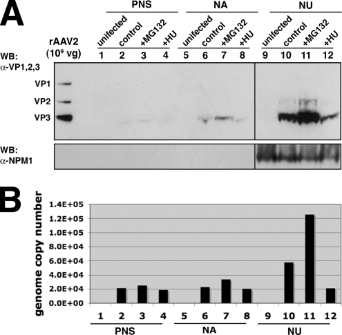
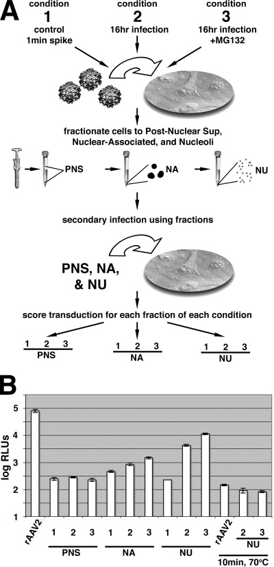
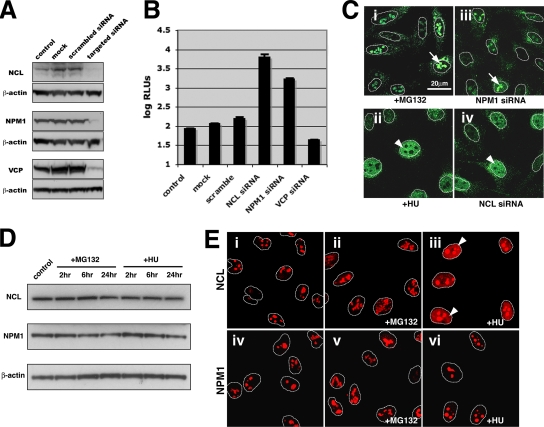
Adeno-associated virus (AAV) serotypes are being tailored for numerous therapeutic applications, but the parameters governing the subcellular fate of even the most highly characterized serotype, AAV2, remain unclear. To understand how cellular conditions control capsid trafficking, we have tracked the subcellular fate of recombinant AAV2 (rAAV2) vectors using confocal immunofluorescence, three-dimensional infection analysis, and subcellular fractionation. Here we report that a population of rAAV2 virions enters the nucleus and accumulates in the nucleolus after infection, whereas empty capsids are excluded from nuclear entry. Remarkably, after subcellular fractionation, virions accumulating in nucleoli were found to retain infectivity in secondary infections. Proteasome inhibitors known to enhance transduction were found to potentiate nucleolar accumulation. In contrast, hydroxyurea, which also increases transduction, mobilized virions into the nucleoplasm, suggesting that two separate pathways influence vector delivery in the nucleus. Using a small interfering RNA (siRNA) approach, we then evaluated whether nucleolar proteins B23/nucleophosmin and nucleolin, previously shown to interact with AAV2 capsids, affect trafficking and transduction efficiency. Similar to effects observed with proteasome inhibition, siRNA-mediated knockdown of nucleophosmin potentiated nucleolar accumulation and increased transduction 5- to 15-fold. Parallel to effects from hydroxyurea, knockdown of nucleolin mobilized capsids to the nucleoplasm and increased transduction 10- to 30-fold. Moreover, affecting both pathways simultaneously using drug and siRNA combinations was synergistic and increased transduction over 50-fold. Taken together, these results support the hypothesis that rAAV2 virions enter the nucleus intact and can be sequestered in the nucleolus in stable form. Mobilization from the nucleolus to nucleoplasmic sites likely permits uncoating and subsequent gene expression or genome degradation. In summary, with these studies we have refined our understanding of AAV2 trafficking dynamics and have identified cellular parameters that mobilize virions in the nucleus and significantly influence AAV infection.


Figures

FIG. 1.
FIG. 1.
Trafficking and transduction after treatment with MG132 or HU. (A) Confocal immunofluorescence microscopy was used to visualize subcellular trafficking of intact virions in HeLa cells with MAb A20 (yellow) with reference to the nuclear membrane marker lamin B1 (magenta). rAAV2 virions (20,000 vg/cell) were administered for the indicated times under control conditions, in the presence of MG132 (2 μM), or following HU pretreatment (10 mM, 12 h). Nucleolar accumulation and diffuse nucleoplasmic localization are marked with white arrows and arrowheads, respectively. The scale bar represents 20 μm. (B) Luciferase assays of transduction were performed to quantify efficiency of firefly luciferase transgene delivery. Two hundred, 2,000, or 20,000 vg/cell was administered for 2 h at 37°C, and cells were washed three times with cell medium, harvested at the indicated times, and scored for transduction after 24 h. RLUs, relative light units.
FIG. 2.
FIG. 2.
Accumulation in the nucleolus is unique to rAAV2. (A) Confocal z-stack analysis of infection was used to determine if the immunofluorescent signal originated from within the nucleus. Serial cross-sections were captured in the z-plane through HeLa cells infected for 16 h in the presence of MG132 (2 μM) with either rAAV2 (i) (20,000 vg/cell) or empty capsids (ii) (equivalent amount relative to A20 and B1 staining). Intact rAAV2 virions (yellow) were found to colocalize with nucleolar marker NCL (magenta) and are depicted within the nucleus (blue, DAPI). (B) Subcellular fractionation of HeLa cells after infection (2,000 vg/cell rAAV2 or equivalent empty capsid amount). Capsid proteins present in the PNS, NA, and NU cell fractions were detected by Western blotting (WB) (MAb B1). The PNS and NA fractions were diluted 25-fold over the NU fraction. Nucleolar isolation was verified by the presence of NPM1 by Western blotting and observing purified nucleoli as dense retractile bodies by phase-contrast microscopy (not shown). (C) Three-dimensional rendering of infection in panel A, as processed by Volocity software (Improvision). The nucleus (blue channel) was gated to reveal nucleoplasmic staining of NCL (magenta) (ii and vi), focal nucleoli (iii and vii), and then the presence or absence of rAAV2 and empty capsids within nucleoli (yellow) (iv and viii). White arrows depict nucleolar accumulation.
FIG. 3.
FIG. 3.
Detection of capsid proteins and viral genomes in nucleoli after subcellular fractionation. (A) Subcellular fractionation of HeLa cells after no infection (lanes 1, 5, and 9), infection with rAAV2 at 2,000 vg/cell (lanes 2, 6, and 10), rAAV2 with Mg132 (lanes 3, 7, and 11), or rAAV2 after HU treatment (lanes 4, 8, and 12). Cells were fractionated after 16 h into PNS, NA, and NU fractions. Capsid proteins in fractions were detected by Western blotting (WB) (MAb B1). Nucleolar isolation is indicated by the presence of NPM1 in the NU fraction. (B) Quantification of viral genomes present in cell fractions after infection by quantitative PCR (same sample order as for panel A). Amplification was performed using Sybr green with primers designed for the luciferase transgene.
FIG. 4.
FIG. 4.
Secondary infection after nucleolar fractionation. (A) Experimental schematic representing three conditions of infection: 1, control with virus present for only 1 min in the cell medium; 2, 16 h of infection with rAAV2 (2,000 vg/cell); and 3, 16 h infection in the presence of MG132 (2 μM). Cells treated in this manner were processed to separate the PNS, NA, and NU fractions. (B) To determine if virions accumulating in these fractions remained infectious, a luciferase assay of transduction was performed 24 h after 5-μl aliquots from the designated fractions were administered to fresh HeLa cells. Particle numbers in samples NU-2 and NU-3 were determined to be similar to those of purified rAAV2 (first bar, 10,000 vg/cell), with NU-3 being slightly higher than NU-2. As a control, purified rAAV2 (10,000 vg/cell) and samples NU-2 and NU-3 were subjected to heat treatment (70°C, 10 min) to destroy capsid integrity. RLUs, relative light units.
FIG. 5.
FIG. 5.
MG132 and HU increase transduction through different mechanisms. (A) Genome pulse-chase experiments were performed to determine whether MG132 or HU influences total levels of vector genome accumulation and to examine the effects of these drugs on genome degradation. Cells were pulsed with rAAV2 (2,000 vg/cell) for 2 h at 37°C, washed with medium, and harvested at 0, 12, and 24 h postinfection. Viral DNA was purified from the cell suspensions and detected by dot blot hybridization using a radiolabeled probe created against the luciferase transgene. (B) A luciferase assay of transduction with rAAV2 (2,000 vg/cell) was performed to test for synergistic transduction enhancement of MG132 and HU. Cells were treated with either HU (10 mM), MG132 (1 μM), or a combination of the two and tested for luciferase activity at 24 h postinfection. RLUs, relative light units.
FIG. 6.
FIG. 6.
Impact of nucleolar proteins on rAAV2 transduction and trafficking. siRNAs targeting NCL and NPM1, nucleolar proteins known to bind AAV capsids, were transfected into HeLa cells at a final concentration of 5 nM in parallel with mock-transfected cells (treated with transfection reagent only), a scrambled siRNA transfection, or siRNA targeting a nonrelated protein, VCP. (A) Western blots of cell lysates taken 4 days after siRNA transfection, detecting loss of targeted protein relative to β-actin controls. (B) Luciferase assay of transduction in HeLa cells transfected with siRNAs targeting NCL, NPM1, or VCP. Cells were split to equal cell densities at 48 h after transfection and infected with rAAV2 (2,000 vg/cell). Transduction was scored 24 h after infection. RLUs, relative light units. (C) Immunofluorescence of rAAV2 in HeLa cells at 16 h postinfection using MAb A20 to detect intact capsids (green), with nuclear membranes outlined relative to DAPI stain (not shown). Patterns of localization were compared between samples treated with MG132 and NPM1 siRNA or samples treated with HU and NCL siRNA. Arrows and arrowheads indicate nucleolar accumulation and diffuse nucleoplasmic staining, respectively. (D) Western blots of total cell protein from control samples or following MG132 or HU administration for 2, 6, or 24 h. Levels of NCL or NPM1 remain relatively unaffected after drug treatments compared to β-actin controls. (E) Immunofluorescence localization of NCL (i, ii, and iii) or NPM1 (iv, v, and vi), shown in red, in untreated cells compared to drug-treated cells after 24 h (MG132, ii and v; HU, iii and vi). Arrowheads indicate a shift toward nucleoplasmic localization of NCL following HU treatment (iii).
FIG. 7.
FIG. 7.
Specific combinations of drugs and siRNAs enhance transduction. HeLa cells transfected with siRNAs targeting NCL, NPM1, or VCP were split to equal cell densities at 48 h after transfection and infected with rAAV2, rAAV2 plus MG132, or rAAV2 plus HU. Transduction efficiency was scored by luciferase assay 24 h later. Asterisks indicate mitigated effectiveness of MG132 in NPM1 siRNA samples, and double asterisks highlight a pronounced reduction in sensitivity to HU in NCL siRNA samples. RLUs, relative light units.
FIG. 8.
FIG. 8.
Illustrated model of two nuclear trafficking paradigms. Virions entering the nucleus are subject to an accumulation pathway (A) and a mobilization pathway (B). Low or negligible transduction occurs when virions do not enter nuclei at high efficiency. If high particle numbers are administered, virions can accumulate in the nucleolus in stable form (A), and favorable conditions may permit vector mobilization within the nucleus to sites that promote uncoating in the nucleoplasm (B). Transduction can be increased by manipulating either pathway independently; however, transduction is dramatically potentiated if more than one pathway is engaged, as exemplified by the cooperative effects of MG132 and HU (Fig. 5B).

Similar articles

Cited by

References

    1. Akache, B., D. Grimm, X. Shen, S. Fuess, S. R. Yant, D. S. Glazer, J. Park, and M. A. Kay. 2007. A two-hybrid screen identifies cathepsins B and L as uncoating factors for adeno-associated virus 2 and 8. Mol. Ther. 15330-339. - PMC - PubMed
    1. Atchison, R. W., B. C. Casto, and W. M. Hammon. 1966. Electron microscopy of adenovirus-associated virus (AAV) in cell cultures. Virology 29353-357. - PubMed
    1. Bantel-Schaal, U., B. Hub, and J. Kartenbeck. 2002. Endocytosis of adeno-associated virus type 5 leads to accumulation of virus particles in the Golgi compartment. J. Virol. 762340-2349. - PMC - PubMed
    1. Barrijal, S., M. Perros, Z. Gu, B. L. Avalosse, P. Belenguer, F. Amalric, and J. Rommelaere. 1992. Nucleolin forms a specific complex with a fragment of the viral (minus) strand of minute virus of mice DNA. Nucleic Acids Res. 205053-5060. - PMC - PubMed
    1. Bartlett, J. S., R. Wilcher, and R. J. Samulski. 2000. Infectious entry pathway of adeno-associated virus and adeno-associated virus vectors. J. Virol. 742777-2785. - PMC - PubMed

Publication types

LinkOut - more resources