. 2009 Mar;83(5):2374-81.
doi: 10.1128/JVI.01898-08. Epub 2008 Dec 24.

Regulation of APOBEC3 proteins by a novel YXXL motif in human immunodeficiency virus type 1 Vif and simian immunodeficiency virus SIVagm Vif

Affiliations

Regulation of APOBEC3 proteins by a novel YXXL motif in human immunodeficiency virus type 1 Vif and simian immunodeficiency virus SIVagm Vif

Erez Pery et al. J Virol. 2009 Mar.

Abstract

The APOBEC3 cytidine deaminases are potent antiviral factors that restrict the replication of human immunodeficiency virus type 1 (HIV-1). In HIV-1-infected CD4+ T cells, the viral accessory protein Vif binds to APOBEC3G (A3G), APOBEC3F (A3F), and APOBEC3C (A3C) and targets these proteins for polyubiquitination by forming an E3 ubiquitin ligase with cullin 5. Previous studies identified regions of HIV-1 Vif, 40YRHHY44 and 12QVDRMR17, which are important for interaction with A3G and A3F, respectively, and showed that Vif residues 54 to 71 are sufficient for A3G binding. Here, we identify 69YXXL72 as a novel conserved motif in HIV-1 Vif that mediates binding to human A3G and its subsequent degradation. Studies on other APOBEC3 proteins revealed that Tyr69 and Leu72 are important for the degradation of A3F and A3C as well. Similar to A3F, A3C regulation is also mediated by Vif residues 12QVDRMR17. Simian immunodeficiency virus (SIV) Vif was shown to bind and degrade African green monkey A3G (agmA3G) and, unexpectedly, human A3C. The YXXL motif of SIVagm Vif was important for the inactivation of agmA3G and human A3C. Unlike HIV-1 Vif, however, SIVagm Vif does not require Tyr40 and His43 for agmA3G degradation. Tyr69 in the YXXL motif was critical for binding of recombinant glutathione S-transferase-Vif(1-94) to A3G in vitro. These results suggest that the YXXL motif in Vif is a potential target for small-molecule inhibitors to block Vif interaction with A3G, A3F, and A3C, and thereby protect cells against HIV-1 infection.


Figures
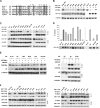
FIG. 1.
FIG. 1.
A conserved YXXL motif in HIV-1 Vif is important for binding and neutralization of hA3G. (A) Sequence alignment of residues 35 to 73 of primate lentivirus Vif proteins. Identical and similar residues are highlighted in dark or light gray, respectively. Vif residues examined for their potential role in A3G neutralization are marked with an asterisk. (B) hA3G-Myc was expressed in 293T cells with wild-type and mutant Vif proteins. Expressed protein levels were determined by Western blot analysis with anti-Myc monoclonal, rabbit anti-Vif, or anti-β-tubulin antibodies. (C) 293T cells were cotransfected with hA3G-HA and wild-type or mutant HIV-1 Vif expression plasmids. Lysates were immunoprecipitated with anti-HA antibody and probed by Western blotting. Equivalent expression levels were confirmed by Western blot analysis of cell lysates. (D) Single-cycle viruses were produced from 293/A3G cells following transfection with a vif-deleted proviral plasmid (pNLX ΔEnv ΔVif-luc), vesicular stomatitis virus glycoprotein (VSVG), and wild-type or mutant pCDNA3.Vif expression plasmids. Infectivity of normalized virus was measured in Cf2 cells. Shown are the means of the results ± the standard deviation (n = 3). In the bottom panel, immunoblots show the expression level of wild-type and mutant HIV-1 Vif proteins in the corresponding producer cells. (E) The W70A Vif mutant induces A3G degradation via the proteasome pathway. Left panel: hA3G-Myc was expressed in 293T cells with wild-type or mutant Vif proteins, and transfected cells were incubated with or without 2.5 μM MG132 for the last 15 h of transfection. Expressed protein levels were determined by Western blot analysis. Right panel: 293T cells were cotransfected with hA3G-HA and wild-type or W70A mutant HIV-1 Vif expression plasmids, and cells were incubated with or without MG132, as in the left panel. Lysates were immunoprecipitated with anti-HA and probed by Western blotting of cell lysates. (F) Mutant HIV-1 Vif proteins bind to Cul5 and EloC. Wild-type or mutant HIV-1 Vif expression plasmids were cotransfected with pCDNA3.HA-Cul5 or pCDNA3.T7-EloC into 293T cells. Lysates were immunoprecipitated with antibodies recognizing epitope tags on the indicated proteins and were probed by Western blotting. Equivalent levels of protein expression were confirmed by Western blotting of cell lysates. WT, wild type; +, present; −, without.
FIG. 2.
FIG. 2.
Tyr69 and Leu72 of HIV-1 Vif are important for degradation of hA3F. (A) hA3F-V5 was expressed in 293T cells with wild-type or mutant HIV-1 Vif proteins. Expressed protein levels were determined by Western blot analysis with anti-V5 monoclonal, rabbit anti-Vif, or anti-β-tubulin antibodies. (B) HIV-1 Vif Tyr69 and Leu72 are important for interaction with hA3F. 293T cells were cotransfected with HA-hA3F and wild-type or mutant HIV-1 Vif plasmids. Lysates were immunoprecipitated with anti-HA antibody and probed by Western blotting. Equivalent levels of protein expression were confirmed by Western blotting of cell lysates. WT, wild type; +, with; −, without.
FIG. 3.
FIG. 3.
YXXL motif in SIVagm Vif is important for neutralization of agmA3G. (A) Sequence alignment of residues 35 to 73 of primate lentivirus Vif proteins from HIV-1 HXB2, SIVagm, and SIVmnd2. Identical and similar residues are highlighted in dark or light gray, respectively. SIVagm Vif residues examined for their ability to neutralize agmA3G are marked with asterisks. (B) Tyr71 and Leu74 residues in SIVagm Vif are important for agmA3G degradation. agmA3G-Myc was expressed in 293T cells with wild-type or mutant SIVagm Vif proteins. Expressed protein levels were determined by Western blotting. (C) Single-cycle viruses were produced from 293T cells following cotransfection with a proviral plasmid (pNLX ΔEnv ΔVif-luc) and plasmids that express VSVG, agmA3G, and wild-type or mutant SIVagmVif proteins. Infectivity of normalized virus was measured in Cf2 cells. Shown are the means of the results ± the standard deviation (n = 3). The bottom panel represents immunoblots that show expression levels of agmA3G and wild-type or mutant HIV-1 Vif proteins in the corresponding producer cells. WT, wild type; +, with; −, without.
FIG. 4.
FIG. 4.
Tyr and Leu residues of the conserved YXXL motif in HIV-1 and SIVagm Vifs are important for hA3C regulation. hA3C-V5 was expressed in 293T cells with wild-type or mutant HIV-1 Vif (A) or SIVagm Vif (B) proteins. Expressed protein levels were determined by Western blot analysis. WT, wild type; +, with; −, without.
FIG. 5.
FIG. 5.
Tyr69 and Trp70 residues in the YXXL motif in recombinant HIV-1 GST-Vif(1-94) are required for binding to hA3G in vitro. (A) A total of 2.5 μg of recombinant GST or wild-type or mutant GST-Vif(1-94) proteins were prebound to glutathione-Sepharose beads. Recombinant His-A3G (200 ng; Immunodiagnostics, Woburn, MA) was added to the prebound beads and incubated overnight at 4°C. Bound GST, GST-Vif, and His-A3G were detected by Western blotting with anti-GST and anti-A3G antibodies. (B) Peptide inhibition of binding between GST-Vif(1-94)-A3G in a homogenous FRET assay (Lance). 15 nM recombinant GST-Vif(1-94) was incubated with 500 nM biotinylated A3G peptide (amino acids 110 to 148) in a 384-well plate. Binding was detected using allophycocyanin-streptavidin and europium-anti-GST antibody and expressed as a FRET ratio [(fluorescence emission intensity at 665 nm/intensity at 615 nm) × 104]. Peptide competition was carried out by adding 2% dimethyl sulfoxide (DMSO) or Vif 15-mer peptides P15 (amino acids 57 to 71) or mutant P15 (amino acids 57 to 71; Y69A/W70A) to Vif-A3G binding reactions at the indicated concentration. Data are presented as means of the results for triplicate samples ± the standard deviations.

References

    1. Bishop, K. N., R. K. Holmes, and M. H. Malim. 2006. Antiviral potency of APOBEC proteins does not correlate with cytidine deamination. J. Virol. 808450-8458. - PMC - PubMed
    1. Bogerd, H. P., B. P. Doehle, H. L. Wiegand, and B. R. Cullen. 2004. A single amino acid difference in the host APOBEC3G protein controls the primate species specificity of HIV type 1 virion infectivity factor. Proc. Natl. Acad. Sci. USA 1013770-3774. - PMC - PubMed
    1. Bourara, K., T. J. Liegler, and R. M. Grant. 2007. Target cell APOBEC3C can induce limited G-to-A mutation in HIV-1. PLoS Pathog. 31477-1485. - PMC - PubMed
    1. Chen, C., F. Li, and R. C. Montelaro. 2001. Functional roles of equine infectious anemia virus Gag p9 in viral budding and infection. J. Virol. 759762-9770. - PMC - PubMed
    1. Chiu, Y. L., V. B. Soros, J. F. Kreisberg, K. Stopak, W. Yonemoto, and W. C. Greene. 2005. Cellular APOBEC3G restricts HIV-1 infection in resting CD4+ T cells. Nature 435108-114. - PubMed

Publication types

Substances