. 2009 May;23(5):1541-57.
doi: 10.1096/fj.08-122184. Epub 2008 Dec 24.

Proteomic and immunologic analyses of brain tumor exosomes

Affiliations

Proteomic and immunologic analyses of brain tumor exosomes

Michael W Graner et al. FASEB J. 2009 May.

Abstract

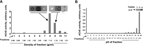
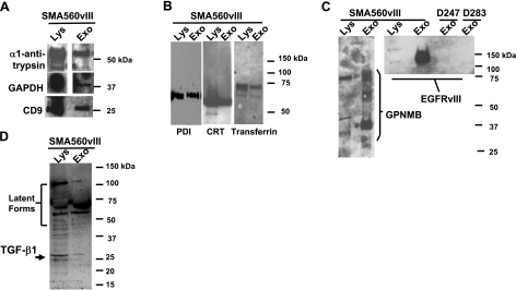
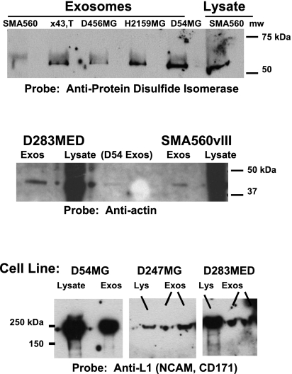
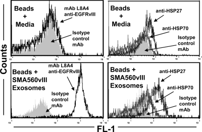
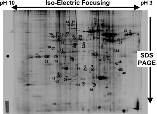
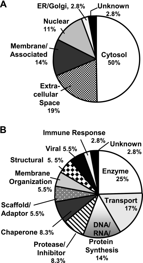
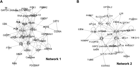
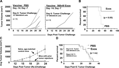
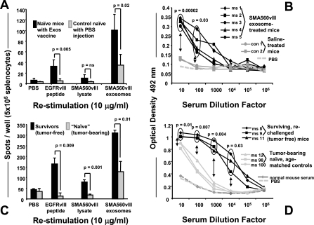
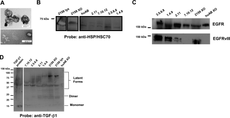
Brain tumors are horrific diseases with almost universally fatal outcomes; new therapeutics are desperately needed and will come from improved understandings of glioma biology. Exosomes are endosomally derived 30-100 nm membranous vesicles released from many cell types into the extracellular milieu; surprisingly, exosomes are virtually unstudied in neuro-oncology. These microvesicles were used as vaccines in other tumor settings, but their immunological significance is unevaluated in brain tumors. Our purpose here is to report the initial biochemical, proteomic, and immunological studies on murine brain tumor exosomes, following known procedures to isolate exosomes. Our findings show that these vesicles have biophysical characteristics and proteomic profiles similar to exosomes from other cell types but that brain tumor exosomes have unique features (e.g., very basic isoelectric points, expressing the mutated tumor antigen EGFRvIII and the putatively immunosuppressive cytokine TGF-beta). Administration of such exosomes into syngeneic animals produced both humoral and cellular immune responses in immunized hosts capable of rejecting subsequent tumor challenges but failed to prolong survival in established orthotopic models. Control animals received saline or cell lysate vaccines and showed no antitumor responses. Exosomes and microvesicles isolated from sera of patients with brain tumors also possess EGFR, EGFRvIII, and TGF-beta. We conclude that exosomes released from brain tumor cells are biochemically/biophysically like other exosomes and have immune-modulating properties. They can escape the blood-brain barrier, with potential systemic and distal signaling and immune consequences.


Figures

Figure 1.
Figure 1.
Characterization of murine brain tumor exosomes by density gradient centrifugation, electron microscopy, free-solution isoelectric focusing and marker (AChE, ALIX) content. A) SMA560vIII exosomes were harvested from spent media by differential centrifugation, followed by density gradient centrifugation. Fractions containing exosomes were identified by AChE activity and negative-stain electron microscopy. Scale bar = 100 nm. B) Exosomes isolated from the appropriate fractions in A were subjected to solution phase isoelectric focusing in a pH 3–10 gradient. AChE activity was used again to identify exosome-containing fractions, and their presence was verified by Western blot analysis with antibodies to ALIX (shown for fractions 19 and 20, with lysate (Lys, 5 μg) as a positive control.
Figure 2.
Figure 2.
Western blot characterization of SMA560vIII exosomes. Exosome proteins (Exo) and lysate proteins (Lys) of parent cells were separated on LDS-PAGE and electroblotted to nitrocellulose. Blots were probed with antibodies against proteins listed. A) Typical proteins found in exosomes: α1-antitrypsin, GAPDH, and the tetraspanin CD9; 20 μg of lysate was loaded. B) Metal binding proteins and chaperones: PDI, CRT, and transferrin; 5 μg of lysate was loaded. C) Known brain tumor antigens: GPNMB and EGFRvIII; 10 μg of lysate was loaded. D) Immunomodulatory cytokine TGF-β1; 10 μg of lysate was loaded. Molecular mass markers for each section are shown at right.
Figure 3.
Figure 3.
Exosomes isolated from a variety of brain tumor cell lines contain the chaperone PDI, the structural protein actin, and the cell adhesion molecule (and brain tumor antigen) L1-NCAM (CD171). Top: exosomes derived from the murine brain tumor model SMA560; from gliomas X43,T, D456MG, and H2159MG (pediatric gliomas); and D54MG all react with anti-PDI antibodies in Western blots. SMA560 lysate is shown as a positive control; 5 μg of lysate was loaded. Middle: Western blots for actin in exosome proteins (and lysates) of D283 medulloblastoma, D54MG, and SMA560vIII; 10 μg of lysate was loaded. Bottom: Western blots of lysates and exosomes from D54MG, D247MG, and D283MED are positive for L1-NCAM/CD171, which is considered an operationally specific tumor antigen; 10 μg of lysate was loaded.
Figure 4.
Figure 4.
Brain tumor exosome surfaces display EGFRvIII and heat shock proteins 27 and 70. Exosomes were bound to aldehyde-sulfate latex beads, were stained with fluorescently labeled antibodies to EGFRvIII (mAb L8A4; left panels) and to HSPs 27 and 70 (right panels), and were analyzed by flow cytometry. Staining of beads soaked in cell culture media as controls are shown in top panels. Isotype control staining profiles are shown as gray fill.
Figure 5.
Figure 5.
Two-dimensional polyacrylamide gel electrophoresis of brain tumor exosomes. 2-D PAGE was performed to separate exosome proteins by isoelectric focusing in the first dimension, and by SDS-PAGE in the second dimension. Gel was stained with Brilliant-Blue Coomassie dye; circled and numbered proteins were identified by mass spectrometry (see Table 1).
Figure 6.
Figure 6.
Categorization of proteins identified in Fig. 5 and Table 1 by subcellular and extracellular localization (A) and by putative function (B). A) Percentage determinations are given: cytosolic proteins, 18 of 36 total; secreted in the extracellular space, 7 of 36; membrane and/or membrane associated (both cytoplasmic and extracellular faces) 5 of 36; nuclear, 4 of 36; ER and Golgi, and unknown, 1 of 36 for each. B) Percentage determinations are based on enzymes and metabolic proteins, 9 of 36 total; transport proteins, 6 of 36; DNA- and RNA-binding proteins and translation and protein synthesis, 5 of 36; chaperones and proteases or protease inhibitors, 3 of 36, respectively; scaffold and adaptor proteins, membrane-organizing proteins, structural proteins, and viral proteins, 2 of 36 for each category; immune response and unknown, 1 of 36 for each. In circumstances in which a protein may have multiple functions or localizations, it received assignment based on the preponderance of literature references.
Figure 7.
Figure 7.
Network analyses of brain tumor exosome proteins identified by proteomics and Western blot analysis. Eight networks were discerned using Ingenuity software (Table 2), with the two significant-scoring ones shown here. Proteins derived empirically from this study are shown as gray-filled symbols; direct protein-protein interactions are shown as solid lines; indirect interactions are shown as broken lines. A) Network 1 had pathways in “hematological, immunological, and respiratory disease.” B) Network 2 pathways were involved in “cell-cell signaling/interaction, cancer, and hematological system development and function.”
Figure 8.
Figure 8.
Antitumor responses of mice immunized with SMA560vIII exosomes. A) Groups of 5 VM/Dk mice were immunized with 50 μg of SMA560vIII exosomes (exos) or with saline (PBS) 14 and 7 days prior to subcutaneous SMA560vIII tumor challenge (106 cells). B, C) Individual tumor volumes were monitored over time, as was survival (B), which was statistically significant in a Kaplan-Meier analysis. Prophylactically immunized mice that survived their initial tumor challenge were rechallenged 135 days after the initial tumor inoculation, along with age-matched naive controls (C), and tumor volumes were monitored (shown here as averages). D) In a preestablished orthotopic model, groups of 10 mice were implanted with 5000 SMA560vIII cells intracranially. Four and 11 days later, mice were treated with 50 μg of SMA560vIII exosomes or with PBS, and survival was monitored. Survival differences between the treated and control groups were not significant (ns).
Figure 9.
Figure 9.
ELISPOT and ELISA measurements of immune responses from exosome-immunized mice. A, B) Naive VM/Dk mice were twice immunized with 50 μg of SMA560vIII exosomes (days −14 and −7) or with PBS as controls. On day 0, spleens and sera were harvested and restimulated (as shown) in vitro in ELISPOT assays for INF-γ release from splenocytes (A) and serum antibody titers against 10 μg/well SMA560vIII exosomes in ELISA assays (B). C, D) Same assays as in A and B, except that mice either were survivors of primary and secondary subcutaneous tumor inoculations (following exosome vaccination), or were age-matched controls that were growing tumors at the time of tissue and sera harvest. Statistically significant differences in outputs are noted in the figure.
Figure 10.
Figure 10.
Tumor-derived exosomes and microvesicles are present in sera from patients with high-grade gliomas. Pooled sera from patients with high-grade gliomas were subjected to differential centrifugation as described in Materials and Methods. A) High-speed pellets were sectioned and visualized by electron microscopy (scale bar=100 nm), or were resuspended in SDS-PAGE sample buffer with 1% saponin, and boiled for 15 min. BD) Maximum volumes of exosome samples were loaded (number-decimal samples are patient-derived; huAB refers to exosomes from human AB serum; 2159XO refers to exosomes from H2159MG cells; 2159 lys refers to lysate from those cells, 5 μg loaded). Proteins were separated on 10% Tris-glycine gels (B, C) or 4–20% gels (D) and blotted to nitrocellulose. Blots were probed with antibodies to canonical HSP/HSC70 (B), to EGFR (C, top), to EGFRvIII (mAb L8A4; C, bottom, and to TGF-β1 (D). Note that exosomes from human AB serum do not react with Abs vs. EGFRvIII or TGF-β1 but do react with Abs vs. EGFR and HSP/HSC70. Native human TGF-β1 protein (5 ng) was used as a positive control (D). Molecular mass markers are shown at left. Lines or boxes around blots indicate differences in exposure time or positioning of blot strips.

References

    1. Stupp R, Mason W P, van den Bent M J, Weller M, Fisher B, Taphoorn M J, Belanger K, Brandes A A, Marosi C, Bogdahn U, Curschmann J, Janzer R C, Ludwin S K, Gorlia T, Allgeier A, Lacombe D, Cairncross J G, Eisenhauer E, Mirimanoff R O. Radiotherapy plus concomitant and adjuvant temozolomide for glioblastoma. N Engl J Med. 2005;352:987–996. - PubMed
    1. Ohgaki H, Kleihues P. Population-based studies on incidence, survival rates, and genetic alterations in astrocytic and oligodendroglial gliomas. J Neuropathol Exp Neurol. 2005;64:479–489. - PubMed
    1. Keller S, Sanderson M P, Stoeck A, Altevogt P. Exosomes: from biogenesis and secretion to biological function. Immunol Lett. 2006;107:102–108. - PubMed
    1. Calzolari A, Raggi C, Deaglio S, Sposi N M, Stafsnes M, Fecchi K, Parolini I, Malavasi F, Peschle C, Sargiacomo M, Testa U. TfR2 localizes in lipid raft domains and is released in exosomes to activate signal transduction along the MAPK pathway. J Cell Sci. 2006;119:4486–4498. - PubMed
    1. Safaei R, Larson B J, Cheng T C, Gibson M A, Otani S, Naerdemann W, Howell S B. Abnormal lysosomal trafficking and enhanced exosomal export of cisplatin in drug-resistant human ovarian carcinoma cells. Mol Cancer Ther. 2005;4:1595–1604. - PubMed

Publication types