. 2009 Feb;20(4):1167-79.
doi: 10.1091/mbc.e08-01-0065. Epub 2008 Dec 24.

Dorsal root ganglion neurons react to semaphorin 3A application through a biphasic response that requires multiple myosin II isoforms

Affiliations

Dorsal root ganglion neurons react to semaphorin 3A application through a biphasic response that requires multiple myosin II isoforms

Jacquelyn A Brown et al. Mol Biol Cell. 2009 Feb.

Abstract

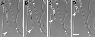
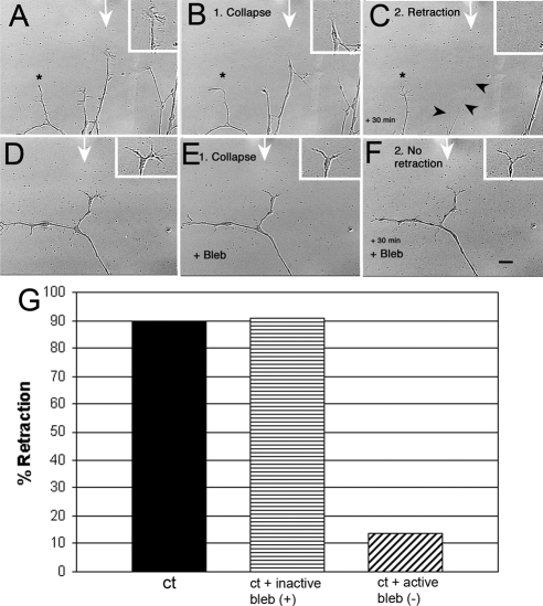
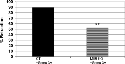
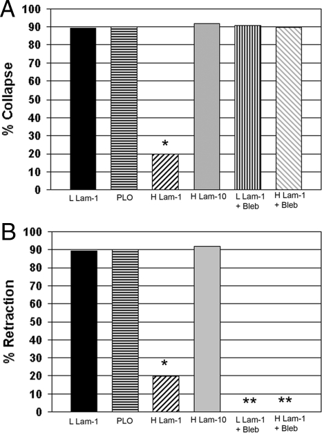
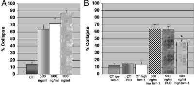
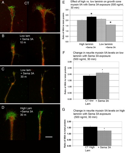
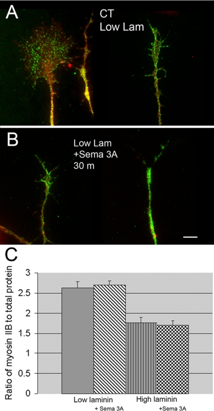
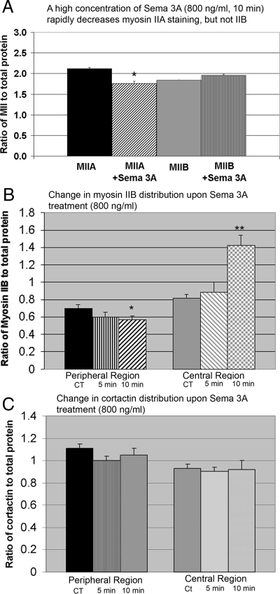
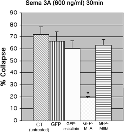
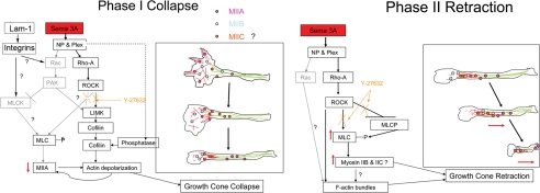
Growth cone responses to guidance cues provide the basis for neuronal pathfinding. Although many cues have been identified, less is known about how signals are translated into the cytoskeletal rearrangements that steer directional changes during pathfinding. Here we show that the response of dorsal root ganglion (DRG) neurons to Semaphorin 3A gradients can be divided into two steps: growth cone collapse and retraction. Collapse is inhibited by overexpression of myosin IIA or growth on high substrate-bound laminin-1. Inhibition of collapse also prevents retractions; however collapse can occur without retraction. Inhibition of myosin II activity with blebbistatin or by using neurons from myosin IIB knockouts inhibits retraction. Collapse is associated with movement of myosin IIA from the growth cone to the neurite. Myosin IIB redistributes from a broad distribution to the rear of the growth cone and neck of the connecting neurite. High substrate-bound laminin-1 prevents or reverses these changes. This suggests a model for the Sema 3A response that involves loss of growth cone myosin IIA to facilitate actin meshwork instability and collapse, followed by myosin IIB concentration at the rear of the cone and neck region where it associates with actin bundles to drive retraction.


Figures

Figure 1.
Figure 1.
Characterization of DRG growth cone responses to bath-applied Sema 3A. (A–D) Time-lapse images showing the three responses observed during bath application of Sema 3A. A growth cone collapses (small white arrowhead) while another retracts (large white arrowhead). A third growth cone extends normally (arrow). Images are at 2-min intervals. Scale bar, 20 μm.
Figure 2.
Figure 2.
DRG neurons for control mice show myosin II–dependent retraction in response to a Sema 3A gradient. Cultures were preincubated with either 100 μm inactive blebbistatin (+) or 10 μm active blebbistatin (−) for 30 min. A gradient of Sema 3A (7.5 μg/ml in the pipette) was applied for 30 min during time-lapse imaging (1-min intervals) via micropipette. The time-lapse movie was used to score for extension, collapse, retraction, and turning. (A–C) A sequence showing typical DRG neurite responses to a Sema 3A gradient when cells are grown on low laminin-1. The micropipette tip was just outside the field at the point indicated by the white arrow. A growth cone in line with the Sema 3A gradient first collapses (15 min) (B) and then retracts (30 min; C). Arrowheads, residual retracted neurite. An adjacent neurite (*) further from the gradient does not retract. (D–F) Blebbistatin (Bleb) treatment does not prevent collapse (E), but does prevent retraction (F). Scale bar, 10 μm. (G) Inactive blebbistatin (+) had no effect on retraction compared with untreated controls, but active blebbistatin (−) caused a significant drop in the number of cells retracting (p = 0.0001; Ct, n = 28; Bleb (−), n = 37; Bleb inactive, n = 11) indicating that myosin II activity is necessary for retraction in response to Sema 3A gradients.
Figure 3.
Figure 3.
Retraction in response to a Sema 3A gradient is partially myosin IIB–dependent. DRG neurons from control type (ct) or myosin IIB knockout (KO) where subjected to a Sema 3A gradient during time-lapse imaging and scored for extension, collapse, or retraction. Myosin IIB knockout neurites showed a significant (36.9%) reduction in retraction frequency (p = 0.0075; Ct, n = 28; KO, n = 25).
Figure 4.
Figure 4.
Increased amounts of substrate-bound laminin-1 can reduce the frequency of collapse and retraction in response to a Sema 3A gradient. Inhibiting myosin II with Bleb only prevents retraction. DRG cultures (normal control-type cells) on coverslips coated with low laminin-1 (9.6 μg/ml), poly-l-ornithine (PLO-0.1 mg/ml), high laminin-1 (32 μg/ml), or high laminin-10 (32 μg/ml) were subjected to a Sema 3A gradient and then scored for collapse (A) and retraction (B). (A) Only high laminin-1 prevented collapse (*p = 0.0001, n = 25 for each condition). Addition of active Bleb to DRG neurons growing on either low laminin-1 or high laminin-1 before exposure to the Sema 3A gradient resulted in collapse. Therefore Bleb treatment eliminated the protective effect of high laminin-1. (B) The same cells were also scored for retraction. High laminin-1 significantly inhibited retraction at the same rate as collapse (*p = 0.0001, n = 25). Active Bleb inhibited retraction for cells growing on both low and high laminin-1. *No retraction was observed in the Bleb-treated cells.
Figure 5.
Figure 5.
Bath applied Sema 3A produces mainly collapse at low concentrations and the response is affected by the amount of substrate-bound laminin-1. (A) The concentration dependence of collapse to bath-applied Sema 3A. DRG cultures were fixed at 30 min of exposure. (B) The amount of substrate-bound laminin-1 has no effect on spontaneous collapse in controls (CT). On exposure to Sema 3A (500 ng/ml) the frequency of collapse on high laminin (*) was significantly reduced compared with low laminin (p = 0.034, n = 4 explants for each condition with a minimum 40 growth cones per explant).
Figure 6.
Figure 6.
Low concentrations of bath-applied Sema 3A decrease growth cone Myosin IIA immunofluorescence staining intensity ratio. High laminin-1 produces the opposite effect. (A) Control (CT) DRG neurons grown on low laminin and then fixed and antibody labeled for myosin IIA (green) and stained for total protein (red). (B) Antibody labeling for myosin IIA and total protein staining after 10 min of Sema 3A treatment (500 ng/ml). The myosin IIA immunofluorescence staining appears more diffuse. Cells were growing on low laminin. (C) Antibody labeling for myosin IIA and staining for total protein after 30 min of Sema 3A treatment. Growth cones have partially collapsed and the myosin IIA staining is decreased. Cells were growing on low laminin-1. (D) Cells labeled for myosin IIA and total protein after 30 min exposure to bath-applied Sema 3A (500 ng/ml). Cells shown were growing on high laminin-1 and did not collapse. Myosin IIA labeling appears similar to control. Scale bar, 5 μm. (E) For quantitative comparisons the antibody staining fluorescence intensity in the growth cone was expressed as a ratio to fluorescence labeling intensity of total protein using activated Cy3 dye. Cells exposed to Sema 3A (500 ng/ml) for 30 min growing on high laminin-1 showed a significant increased in myosin IIA immunofluorescence staining (*p = 0.0001). In contrast cells exposed to Sema 3A (500 ng/ml) for 30 min growing on low laminin-1 showed significantly decreased staining for myosin IIA (*p = 0.0001). For each condition N = 40 growth cones. (F) Myosin IIA staining redistributes within the neurite after Sema 3A exposure. The same cells used for E were analyzed for changes in myosin II staining intensity in the neurite after Sema 3A (500 ng/ml) for 30 min. The ratio of myosin IIA to total protein in the neurite, defined as the segment starting 50 μm behind the growth cone. In cases where growth cones were collapsed this measurement was taken starting 10 μm from the tip of the process. The addition of Sema 3A significantly increased the staining ratio for myosin IIA (*p = 0.044, n = 25). (G) The same analysis was done for cells growing on high laminin. Myosin IIA staining ratio in the neurite showed a significant decrease (*p = 0.0037, n = 40). Thus the changes are the opposite of those shown in E, suggesting movement of myosin IIA between the growth cone and neurite.
Figure 7.
Figure 7.
Low concentrations (500 ng/ml) of bath-applied Sema 3A do not change growth cone myosin IIB immunofluorescence staining intensity ratios. (A) Growth cones for control type DRG neurons growing on low laminin-1 fixed and antibody labeled for myosin IIB (green) and stained for total protein (red). (B) Antibody labeling for myosin IIB and staining for total protein after 30 min of Sema 3A treatment (500 ng/ml). Growth cones have partially collapsed but the myosin IIB staining intensity appears unchanged. Cells were growing on low laminin. The staining results on high laminin were the same except that collapse was inhibited. (C) For quantitative comparisons the antibody staining fluorescence intensity in the growth cone was expressed as a ratio to fluorescence labeling intensity of total protein using activated Cy3 dye. Cells exposed to Sema 3A (500 ng/ml) for 30 min growing on either low or high laminin-1 showed no significant change in the myosin IIB immunofluorescence staining ratios (low laminin p = 0.7, n = 60, high laminin p = 0.6, n = 25). Scale bar, 6 μm.
Figure 8.
Figure 8.
High concentrations of bath-applied Sema 3A induce changes in total growth cone myosin IIA–staining intensity ratio, but not myosin IIB. (A) Exposure of DRG neurons to bath-applied Sema 3A at a high concentration (800 ng/ml) for 10 min induced a significant decrease in myosin IIA–staining ratio. Under the same conditions, the myosin IIB staining ratio did not change. Most growth cones collapse and many neurites retract under these conditions. For fully collapsed neurites the analysis was performed on the last 10 μm. Retracted neurites were not included in the analysis. (B) High concentrations (800 ng/ml) of bath-applied Sema 3A induce redistributions of myosin IIB in the growth cone at short times (5 and 10 min). The myosin IIB staining ratio was analyzed in peripheral and central regions of growth cones. Only noncollapsed growth cones were selected for analysis at the different time points. For comparison the immunofluorescence staining intensity of a specific actin-binding protein (cortactin) was analyzed in the same growth cones (C). Peripheral myosin IIB showed a significant decrease at 10 min (*p = 0.0001, n = 18) and a corresponding significant increase in the central region (*p = 0.03, n = 18). The magnitude of the increase in the central region was greater than the decrease in peripheral region. However the area of the peripheral region is much larger than the central region. (C) In the same growth cones the cortactin staining ratio did not significantly change.
Figure 9.
Figure 9.
Over expression of GFP-myosin IIA, but not GFP-myosin IIB blocks collapse in response to relatively high bath-applied Sema 3A (600 ng/ml, 30 min). DRG neurons were transfected with GFP, GFP-α-actinin, GFP-MIIA, and GFP-MIIB. Cultures were fixed and scored for collapse or imaged by time lapse and fixed before scoring for collapse. Only cells expressing GFP-MIIA showed a significant reduction in collapse frequency (*p ≤ 0.001, in order n = 80, 45, 36, 32, and 49) compared with GFP-expressing cells. An additional experiment gave similar results.
Figure 10.
Figure 10.
A model for the molecular interaction involved in the two phases of the Sema 3A response. Phase 1 is collapse and involves loss of myosin IIA and actin filaments from the growth cone. The loss may be simultaneous or sequential and is likely to lead to a transient decrease in tension on the neurite. Activation of integrins by high substrate-bound laminin-1 can modify the response, but the pathways remain unclear. ROCK appears to be partially involved in controlling collapse (Gallo, 2006), but other pathways may also contribute (indicated in gray). Phase II is retraction and involves a redistribution of myosin IIB (and possibly IIC), as well as an increase in bundled actin at the rear of the growth cone or neck region followed by actomyosin drive contractions to increase tension. If the contractions are sufficiently compressed in time and tension overcomes the adhesive interactions with the substrate, then the neurite fully retracts. ROCK appears to also control this phase, although its interactions are complex and need further work to obtain a complete picture. Other pathways may contribute to this phase.

References

    1. Ahmad F. J., Hughey J., Wittmann T., Hyman A., Greaser M., Baas P. W. Motor proteins regulate force interactions between microtubules and microfilaments in the axon. Nat. Cell Biol. 2000;5:276–280. - PubMed
    1. Allingham J. S., Smith R., Rayment I. The structural basis of blebbistatin inhibition and specificity for myosin II. Nat. Struct. Mol. Biol. 2005;12:378–379. - PubMed
    1. Aizawa H., Wakatsuki S., Isii A., Moriyama K., Sasaki Y., Ohashi K., Sekine-Aizawa Y., Sehara-Fujisawa A., Mizuno K., Goshima Y., Yahara I. Phosphorylation of cofilin by LIM-kinase is necessary for semaphorin 3A-induced growth cone collapse. Nat. Neurosci. 2001;4:367–373. - PubMed
    1. Behar O., Mizuno K., Badminton M., Woolf C. J. Semaphorin 3A growth cone collapse requires a sequence homologous to tarantula hanatoxin. Proc. Natl. Acad. Sci. USA. 1999;96:13501–13505. - PMC - PubMed
    1. Bridgman P. C., Dave S., Asnes C. F., Tullio A. N., Adelstein R. S. Myosin IIB is required for growth cone motility. J. Neurosci. 2001;21:6159–6169. - PMC - PubMed

Publication types

MeSH terms

LinkOut - more resources