. 2009 Jan 6;106(1):157-62.
doi: 10.1073/pnas.0811426106. Epub 2008 Dec 24.

Reprogramming of murine and human somatic cells using a single polycistronic vector

Affiliations

Reprogramming of murine and human somatic cells using a single polycistronic vector

Bryce W Carey et al. Proc Natl Acad Sci U S A. .

Erratum in

  • Proc Natl Acad Sci U S A. 2009 Jul 14;106(28):11818
  • Proc Natl Acad Sci U S A. 2009 Mar 31;106(13):5449

Abstract

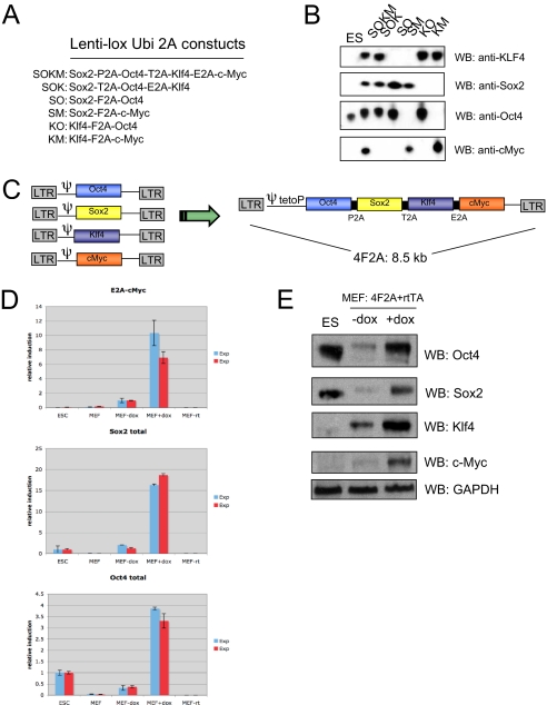
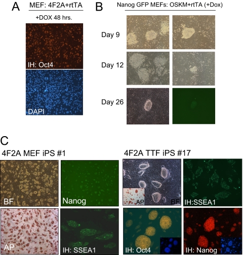
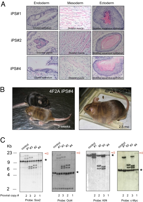
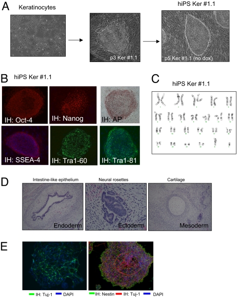
Directed reprogramming of somatic cells by defined factors provides a novel method for the generation of patient-specific stem cells with the potential to bypass both the practical and ethical concerns associated with somatic cell nuclear transfer (SCNT) and human embryonic stem (hES) cells. Although the generation of induced pluripotent stem (iPS) cells has proven a robust technology in mouse and human, a major impediment to the use of iPS cells for therapeutic purposes has been the viral-based delivery of the reprogramming factors because multiple proviral integrations pose the danger of insertional mutagenesis. Here we report a novel approach to reduce the number of viruses necessary to reprogram somatic cells by delivering reprogramming factors in a single virus using 2A "self-cleaving" peptides, which support efficient polycistronic expression from a single promoter. We find that up to four reprogramming factors (Oct4, Sox2, Klf4, and c-Myc) can be expressed from a single virus to generate iPS cells in both embryonic and adult somatic mouse cells and we show that a single proviral copy is sufficient to generate iPS cells from mouse embryonic fibroblasts. In addition we have generated human induced pluripotent stem (hiPS) cell lines from human keratinocytes, demonstrating that a single polycistronic virus can reprogram human somatic cells.


Conflict of interest statement

Conflict of interest statement: R.J. is an advisor to Stemgent.

Figures

Fig. 1.
Fig. 1.
Generation of murine iPS cells using a single 4F2A polycistronic virus. (A) FUW lentivirus constructs tested by transient transfection. In total, four 2A peptides (F2A, T2A, E2A, and P2A) were used. (B) Transient transfection of 293 cells with FUW 2A lentiviruses. Cells were harvested after 48 h and analyzed by Western blot (WB). Efficient protein expression was observed in all constructs tested, indicating four unique 2A peptides support robust protein expression. Note: Sox2 protein is not detected in ES cells because only a short exposure was used. (C) Schematic of the 4F2A DOX-inducible lentivirus containing three types of 2A peptides (P2A, T2A, and E2A). Murine cDNAs for Oct4, Sox2, Klf4, and c-Myc. This particular sequence of factors and 2A peptides is subsequently referred to as “4F2A.” (D) RT-PCR anaylsis of mRNA induction in cells transduced with OSKM 4F2A + rtTA for 3 days. Total Oct4 or Sox2 induction was used to test levels of 4F2A induction relative to ES cells. E2A-cMyc primers were used to detect viral-specific transcripts. Error bars represent SD of the mean of triplicate reactions. (E) Western blot analysis of MEFs transduced with 4F2A + rtTA for 3 days. Cells infected with 4F2A DOX-inducible lentivirus + rtTA produce all four reprogramming factors upon addition of doxycycline (DOX).
Fig. 2.
Fig. 2.
4F2A iPS cells express pluripotency markers. (A) Immunostaining of Oct4 protein indicates high titer infections can be achieved with the 4F2A. MEFs were cultured in DOX media for 2 days after transduction with 4F2A + rtTA. (B) Morphology changes in Nanog-GFP MEFs transduced with 4F2A + rtTA cultured in ES media + DOX. Colonies appeared at ≈8 days, similar to cells infected with single viruses. Nanog GFP+ colonies were observed by day 25 after DOX media removal at day 20. Two columns show typical colonies observed on the plate. (C) 4F2A iPS lines generated from Nanog-GFP MEFs or 14-week tail-tip fibroblasts (TTFs) that stain positive for pluripotency markers AP, SSEA1, Oct4 and have reactivated the endogenous Nanog locus (GFP+ for MEFs and by immunostaining for TTF).
Fig. 3.
Fig. 3.
4F2A iPS cells are pluripotent and contain between 1 and 3 proviral integrations. (A) In vivo differentiation of 4F2A MEF-iPS lines nos. 1, 2, and 4. Histological analysis of teratomas induced after s.c. injection into SCID mice indicates iPS lines contribute to all three germ layers. (B) Moderate-to-high contribution in postnatal chimeric mice as detected by agouti coat color from 4F2A iPS line no. 4. (C) Southern blot analysis of 4F2A proviral integrations in MEF-iPS cell line nos. 1–4. iPS cell DNA was digested with BamHI. Hybridization of the same molecular weight fragment using all four probes indicates presence of 4F2A provirus. Red arrow highlights iPS line no. 4, which contained one proviral copy of the 4F2A. * indicates endogenous allele.
Fig. 4.
Fig. 4.
Generation of human iPS lines using a single 4F2A polycistronic virus. (A) Neonatal human foreskin keritinocytes (NHFK) transduced with 4F2A (carrying mouse cDNAs) + rtTA. On day 22 a single colony was picked and expanded, giving rise to colonies resembling hES colonies. These colonies were picked and a stable hiPS line was established. (B) Ker-hiPS no. 1.1 immunostaining for pluripotency markers AP, Oct4, Nanog, SSEA-4, Tra1–60, and Tra1–81. DAPI stain is in lower panels. (C) Karyotype of Ker-hiPS no. 1.1 is normal 46 XY. (D) In vivo differentiation of Ker-hiPS no. 1.1. Hematoxylin and eosin staining of teratoma sections generated by Ker-hiPS no. 1.1. (E) In vitro differentiation of Ker-hiPS no. 1.1. (Left) Ker-iPS no. 1.1-derived neural precursors exposed to differentiation conditions for 6 days produce terminally differentiated neurons as detected by anti-Tuj1 immunostaining (green). (Right) Ker-iPS no. 1.1 neural precursors (NPs) undergo spontaneous differentiation. NPs were detected by anti-Nestin immunostaining and differentiated neurons by anti-Tuj1 (red). DAPI stain for DNA in both pictures is blue.

References

    1. Lowry WE, et al. Generation of human induced pluripotent stem cells from dermal fibroblasts. Proc Natl Acad Sci USA. 2008;105:2883–2888. - PMC - PubMed
    1. Maherali N, et al. Directly reprogrammed fibroblasts show global epigenetic remodeling and widespread tissue contribution. Cell Stem Cell. 2007;1:55–70. - PubMed
    1. Okita K, Ichisaka T, Yamanaka S. Generation of germline-competent induced pluripotent stem cells. Nature. 2007;448:313–317. - PubMed
    1. Park IH, et al. Reprogramming of human somatic cells to pluripotency with defined factors. Nature. 2008;451:141–146. - PubMed
    1. Takahashi K, et al. Induction of pluripotent stem cells from adult human fibroblasts by defined factors. Cell. 2007;131:861–872. - PubMed

Publication types

Substances