. 2009 Jan 6;106(1):163-8.
doi: 10.1073/pnas.0811631106. Epub 2008 Dec 24.

A role for the host coatomer and KDEL receptor in early vaccinia biogenesis

Affiliations

A role for the host coatomer and KDEL receptor in early vaccinia biogenesis

Leiliang Zhang et al. Proc Natl Acad Sci U S A. .

Abstract

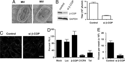
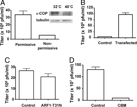
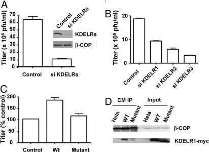
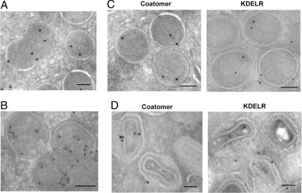
Members of the poxvirus family have been investigated for their applications as vaccines and expression vectors and, more recently, because of concern for their potential as biological weapons. Vaccinia virus, the prototypic member, evolves through multiple forms during its replication. Here, we show a surprising way by which vaccinia hijacks coatomer for early viral biogenesis. Whereas coatomer forms COPI vesicles in the host early secretory system, vaccinia formation bypasses this role of coatomer, but instead, depends on coatomer interacting with the host KDEL receptor. To gain insight into the viral roles of these two host proteins, we have detected them on the earliest recognized viral forms. These findings not only suggest insights into early vaccinia biogenesis but also reveal an alternate mechanism by which coatomer acts.


Conflict of interest statement

The authors declare no conflict of interest.

Figures

Fig. 1.
Fig. 1.
Coatomer acts in vaccinia formation. (A) HeLa cells were infected with the WR strain of vaccinia virus for 16 h and then examined by EM with ImmunoGold labeling for β-COP. (Scale bar: 50 nm.) (B) HeLa cells, either treated with siRNA against β-COP for 48 h or mock-treated, were then infected with WR virus for 24 h. The vaccinia replication assay was then performed, with the mean and standard error from 3 experiments shown (P < 0.01). Gel shows the level of β-COP comparing siRNA-treated versus mock-treated HeLa cells. (C) The surface pool of TfR on HeLa cells, either treated with siRNA or mock-treated, was labeled with fluorescently tagged Tf, and then tracked for internalization into early endosomes. (Scale bar: 10 μm.) (D) TZM-bl cells were transfected with siRNAs against different targets, β-COP, CXCR4, Tat, or Luciferase as control, or mock-treated. After infection with HIV-IIIB, β-galactosidase activity was measured, which was expressed as relative light units (RLU), and then normalized to the condition of mock treatment. The mean with standard deviation from 3 experiments is shown. (E) HeLa cells, either treated with siRNA against β-COP for 48 h or mock-treated, were then infected with WR virus for 24 h. Intracellular viral forms were then purified from infected cells, followed by EM examination for intact viruses. Quantitation was performed from 10 randomly selected images, and then expressed as mean with standard error (P < 0.01).
Fig. 2.
Fig. 2.
Characterizing how coatomer acts in vaccinia formation. (A) A CHO cell line that expressed a temperature-sensitive mutant of ε-COP was incubated either at permissive or nonpermissive temperature for 12 h, and then infected with a recombinant virus (vP30CP77, capable of replicating in CHO cells) for another 24 h. The vaccinia replication assay was then performed, with the mean and standard error from 3 experiments shown (P < 0.01). Gel shows the level of ε-COP, comparing permissive versus nonpermissive temperatures. (B) The mutant CHO cell line that expressed a temperature-sensitive mutant of ε-COP was stably transfected with wild-type ε-COP. After shifting to the nonpermissive temperature for 12 h followed by infection with the recombinant virus (vP30CP77) for 24 h, the vaccinia replication assay was then performed, with the mean and standard error from 3 experiments shown (P < 0.01). (C) HeLa cells, transiently transfected with ARF1-T31N for 48 h or mock-treated, were infected with WR virus for 24 h. The vaccinia replication assay was then performed, with the mean and standard error from 3 experiments shown (P = 0.15). (D) HeLa cells were infected with WR virus for 24 h, with CBM treatment (or mock treatment with vehicle alone) given at the start of the infection. The vaccinia replication assay was then performed, with the mean and standard error from 3 experiments shown (P < 0.01).
Fig. 3.
Fig. 3.
Interaction with the KDELR is important for the viral role of coatomer. (A) HeLa cells, either treated with a combination of siRNAs that targets all human KDELR genes for 48 h or mock-treated, were infected with WR virus for 24 h. The vaccinia replication assay was then performed, with the mean and standard error from 3 experiments shown (P < 0.01). Gel shows protein levels in whole-cell lysate by immunoblotting for proteins indicated. (B) HeLa cells, either treated with siRNAs against individual KDELR genes as indicated for 48 h or mock-treated, were infected with WR virus for 24 h. The vaccinia replication assay was then performed, with the mean and standard error from 3 experiments shown. (C) HeLa cells, stably transfected with either a siRNA-resistant form of wild-type KDELR1 or a mutant KDELR1 defective in binding to coatomer or untransfected as control, were treated with siRNA against native KDELR1 for 48 h. Cells were then infected with WR virus for 24 h. The vaccinia replication assay was then performed to obtain a mean with standard error from 3 experiments. Values are normalized to that of control cells. (D) HeLa cells treated with siRNA against KDELR1, stably expressing wild-type KDELR1, mutant KDELR1, or untransfected as control, were infected with virus for 24 h. Coatomer was then immunoprecipitated from cells from the different conditions followed by immunoblotting for the proteins indicated.
Fig. 4.
Fig. 4.
Distributions of coatomer and the KDELR on early viral forms. (A) HeLa cells were treated with rifampin and then infected with WR virus for 14 h. After washing out rifampin for 30 min, infected cells were examined by EM with ImmunoGold labeling for β-COP. (B) Cells were infected, treated, and then examined in the same manner as described in A, except ImmunoGold labeling was performed for KDELR. (C) Cells were infected, treated, and then examined in the same manner as described in A. ImmunoGold labeling was performed for either β-COP or KDELR. (D) Cells were infected, treated, and then examined in the same manner as described in A. ImmunoGold labeling was performed for either β-COP or KDELR. (Scale bars: 50 nm.)
Fig. 5.
Fig. 5.
KDELR resides in assembling early viral structures that contain lipids. (A) Cells were infected, treated, and then examined in the same manner as described in Fig. 4, with ImmunoGold labeling for either β-COP or KDELR. (Scale bar: 50 nm.) (B) Infected cells subjected to rifampin washout, as described in Fig. 4, were homogenized and then loaded at the bottom of a sucrose density gradient. After equilibrium centrifugation, fractions from the gradient were immunoblotted for KDELR. (C) HeLa cells transiently transfected with the mutant huntingtin protein tagged by GFP were analyzed in a sucrose density gradient as described in B.

References

    1. Condit RC, Moussatche N, Traktman P. In a nutshell: Structure and assembly of the vaccinia virion. Adv Virus Res. 2006;66:31–124. - PubMed
    1. Sodeik B, Krijnse-Locker J. Assembly of vaccinia virus revisited: De novo membrane synthesis or acquisition from the host? Trends Microbiol. 2002;10:15–24. - PubMed
    1. Heuser J. Deep-etch EM reveals that the early poxvirus envelope is a single membrane bilayer stabilized by a geodetic “honeycomb” surface coat. J Cell Biol. 2005;169:269–283. - PMC - PubMed
    1. McMahon HT, Mills IG. COP and clathrin-coated vesicle budding: Different pathways, common approaches. Curr Opin Cell Biol. 2004;16:379–391. - PubMed
    1. Hosobuchi M, Kreis T, Schekman R. SEC21 is a gene required for ER to Golgi protein transport that encodes a subunit of a yeast coatomer. Nature. 1992;360:603–605. - PubMed

Publication types

LinkOut - more resources