. 2008 Dec 24;28(52):14282-92.
doi: 10.1523/JNEUROSCI.4968-08.2008.

Retinal ganglion cells survive and maintain normal dendritic morphology in a mouse model of inherited photoreceptor degeneration

Affiliations

Retinal ganglion cells survive and maintain normal dendritic morphology in a mouse model of inherited photoreceptor degeneration

Francesca Mazzoni et al. J Neurosci. .

Abstract

Retinitis pigmentosa (RP), a family of inherited disorders characterized by progressive photoreceptor death, is a leading cause of blindness with no available cure. Despite the genetic heterogeneity underlying the disease, recent data on animal models show that the degeneration of photoreceptors triggers stereotyped remodeling among their postsynaptic partners. In particular, bipolar and horizontal cells might undergo dendritic atrophy and secondary death. The aim of this study was to investigate whether or not concomitant changes also occur in retinal ganglion cells (RGCs), the only retinal projection neurons to the brain and the proposed substrate for various therapeutic approaches for RP. We assessed the retention of morphology, overall architecture, and survival of RGCs in a mouse model of RP at various stages of the disease. To study the morphology of single RGCs, we generated a new mouse line by crossing Thy1-GFP-M mice (Feng et al., 2000), which express GFP (green fluorescent protein) in a small number of heterogeneous RGCs types, and rd10 mutants, a model of autosomal recessive RP, which exhibit a typical rod-cone degeneration (Chang et al., 2002). We show remarkable preservation of RGC structure, survival, and projections to higher visual centers in the time span from 3 to 9 months of life, well beyond the death of photoreceptors. Thus, unlike second-order neurons, RGCs appear as a considerably stable population of cells, potentially constituting a favorable substrate for restoring vision in RP individuals by means of electronic prostheses or direct expression of photosensitive proteins.


Figures
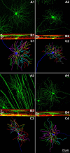
Figure 1.
Figure 1.
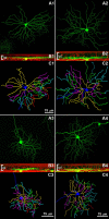
Four types of RGCs of the rd10/Thy1-GFP-M retina with dendritic stratification in the outer one-half of the IPL (functionally, OFF-type RGCs). Age, 9 months. In all RGC illustrations, the top panel (A) represents a whole-mount view of an individual cell projected in a single plane; the central panel (B) is the orthogonal view on the same cells obtained by computer rotation, also showing ethidium nuclear staining (red signal); the bottom panel (C) represents the Neurolucida drawing of each cells. Different colors code for different branching order of the dendrites: blue, first order; yellow, second; orange, third; pink, fourth; green, fifth; cyan, sixth; red, seventh; red, brown, eighth; purple, ninth; white, tenth. A1–C1, GCs belonging to the A2 outer type. A2–C2, GCs belonging to the B1 type. A3–C3, GCs classified as B3 outer type. A4–C4, Example of ganglion cells belonging to the C2 outer type.
Figure 2.
Figure 2.
Four types of RGCs of the rd10/Thy1-GFP-M retina with dendritic stratification in the inner one-half of the IPL (functionally, ON-type RGCs). Age, 9 months. A1–C1, Example of A1 type GC. A2–C2, Example of A2 inner GC. A3–C3, Example of B3 inner GC. A4–C4, Example of C2 inner GC.
Figure 3.
Figure 3.
Indicators of dendritic tree complexity for four types of RGCs with dendrites in the outer one-half of the IPL (OFF RGCs). Parameters from the rd10 mutant (gray columns) are compared with corresponding values obtained from wt mice aged 8–9 months (dark columns). Diagrams in the three rows illustrate dendritic tree area, dendritic tree length, and total number of nodes for the three age groups examined (3, 7, and 9 months). Each column represents average data from four to six cells ± SEM. Statistical analysis with one-way ANOVA shows no significant differences between RGCs of rd10/Thy1-GFP-M and control mice (p > 0.01).
Figure 4.
Figure 4.
Indicators of dendritic tree complexity for four types of RGCs with dendrites in the inner one-half of the IPL (ON RGCs). Parameters from the rd10/Thy1-GFP-M mutant (gray columns) are compared with corresponding values obtained from wt mice aged 8 months (dark columns). Diagrams in the three rows illustrate dendritic tree area, dendritic tree length, and total number of nodes for the three age groups examined (3, 7, and 9 months). Each column represents average data from four to six cells ± SEM. Statistical analysis with one-way ANOVA shows no significant differences between RGCs of rd10/Thy1-GFP-M mice and control mice (p > 0.01).
Figure 5.
Figure 5.
Survival of cells in the GCL of rd10/Thy1-GFP-M mice at 9 months. A, B, Images of the GCL in wt (A) and mutant mice (B). Ethidium nuclear staining and projection on a single plane of the entire GCL thickness. Elongated, bright nuclei belong to blood vessel cells. C, Cellular counting demonstrates that there are no differences in the wt (left, 136,000 ± 2040 cells) and mutant retinas (right, 130,750 ± 5100). The two populations are statistically identical (p > 0.01). D–F, Brn3b immunostaining of cells in the GCL. This transcription factor is localized in the cell nuclei. G, Similar densities of Brn3b-positive cells were found in the three strains of mice at 9 months of age. The three populations are statistically identical (p > 0.01).
Figure 6.
Figure 6.
Anterograde axonal transport of RGCs to central projection areas. A, Schematic diagram showing the ipsilateral and contralateral projections of optic nerve axons to the dorsal part of the lateral geniculate nucleus (dLGN). B, C, Coronal sections of the dLGN from wt (B) and rd10 (C) mutant mice aged 9 months, after injections of cholera toxin conjugated to different fluorophores in the two eyes. Each pair of images shows complementary red/green fields, comprising a large projection from the contralateral eye and a small field, receiving the ipsilateral projection. ipsi, Ipsilateral; contra, contralateral; DX, right side; SX, left side.

References

    1. Alpár A, Palm K, Schierwagen A, Arendt T, Gärtner U. Expression of constitutively active p21H-rasval12 in postmitotic pyramidal neurons results in increased dendritic size and complexity. J Comp Neurol. 2003;467:119–133. - PubMed
    1. Badea TC, Nathans J. Quantitative analysis of neuronal morphologies in the mouse retina visualized by using a genetically directed reporter. J Comp Neurol. 2004;480:331–351. - PubMed
    1. Barhoum R, Martínez-Navarrete G, Corrochano S, Germain F, Fernandez-Sanchez L, de la Rosa EJ, de la Villa P, Cuenca N. Functional and structural modifications during retinal degeneration in the rd10 mouse. Neuroscience. 2008;155:698–713. - PubMed
    1. Bi A, Cui J, Ma YP, Olshevskaya E, Pu M, Dizhoor AM, Pan ZH. Ectopic expression of a microbial-type rhodopsin restores visual responses in mice with photoreceptor degeneration. Neuron. 2006;50:23–33. - PMC - PubMed
    1. Carcieri SM, Jacobs AL, Nirenberg S. Classification of retinal ganglion cells: a statistical approach. J Neurophysiol. 2003;90:1704–1713. - PubMed

Publication types

MeSH terms

LinkOut - more resources