Diagnosis and management of microscopic colitis
- PMID: 19109861
- PMCID: PMC2778111
- DOI: 10.3748/wjg.14.7280
Diagnosis and management of microscopic colitis
Abstract
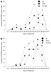
Microscopic colitis, comprising collagenous and lymphocytic colitis, is characterized clinically by chronic watery diarrhea, and a macroscopically normal colonic mucosa where diagnostic histopathological features are seen on microscopic examination. The annual incidence of each disorder is 4-6/100,000 inhabitants, with a peak incidence in 60-70-year-old individuals and a noticeable female predominance for collagenous colitis. The etiology is unknown. Chronic diarrhea, abdominal pain, weight loss, fatigue and fecal incontinence are common symptoms, which impair the health-related quality of life of the patient. There is an association with other autoimmune disorders such as celiac disease, diabetes mellitus, thyroid disorders and arthritis. Budesonide is the best-documented short-term treatment, but the optimal long-term strategy needs further study. The long-term prognosis is good and the risk of complications including colonic cancer is low.
Figures


References
-
- Pardi DS. Microscopic colitis: an update. Inflamm Bowel Dis. 2004;10:860–870. - PubMed
-
- Lindström CG. 'Collagenous colitis' with watery diarrhoea--a new entity? Pathol Eur. 1976;11:87–89. - PubMed
-
- Lazenby AJ, Yardley JH, Giardiello FM, Jessurun J, Bayless TM. Lymphocytic ("microscopic") colitis: a comparative histopathologic study with particular reference to collagenous colitis. Hum Pathol. 1989;20:18–28. - PubMed
Publication types
MeSH terms
Substances
LinkOut - more resources
Full Text Sources
Other Literature Sources

