. 2008 Dec 26;135(7):1311-23.
doi: 10.1016/j.cell.2008.10.044.

Identification of a molecular signaling network that regulates a cellular necrotic cell death pathway

Affiliations

Identification of a molecular signaling network that regulates a cellular necrotic cell death pathway

Junichi Hitomi et al. Cell. .

Abstract

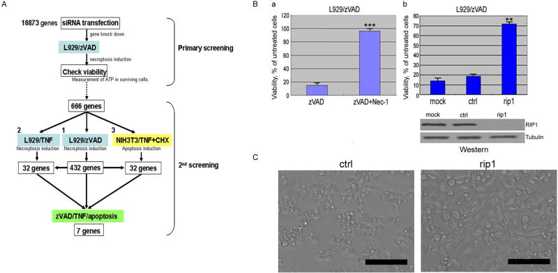
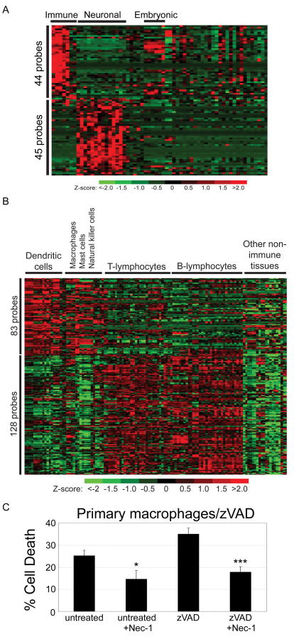
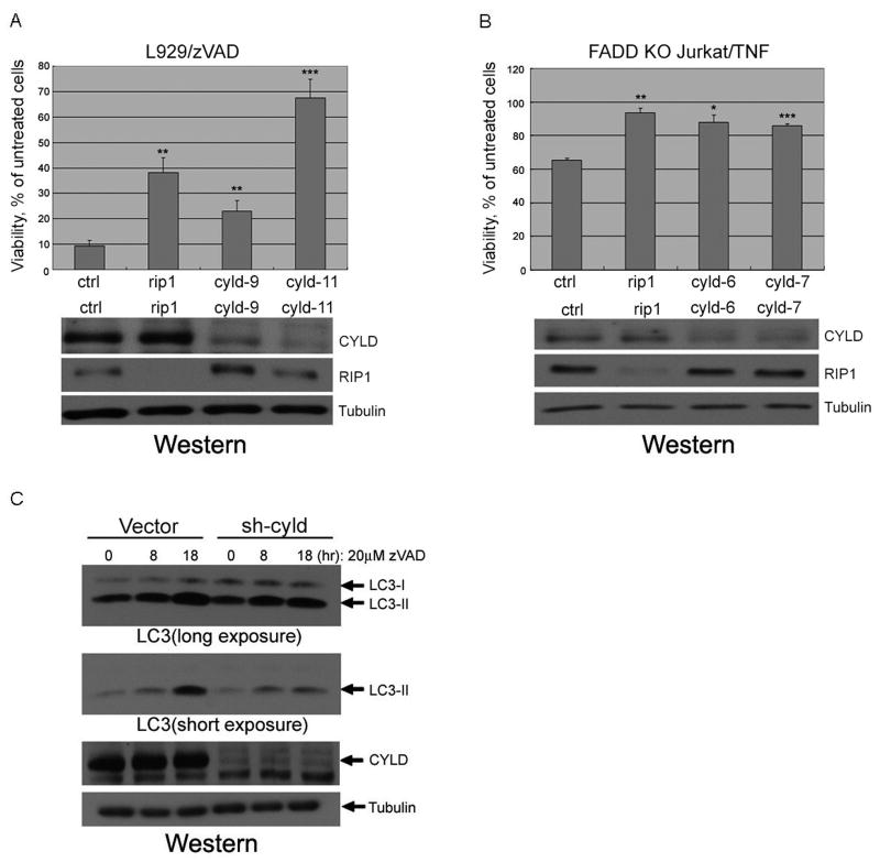
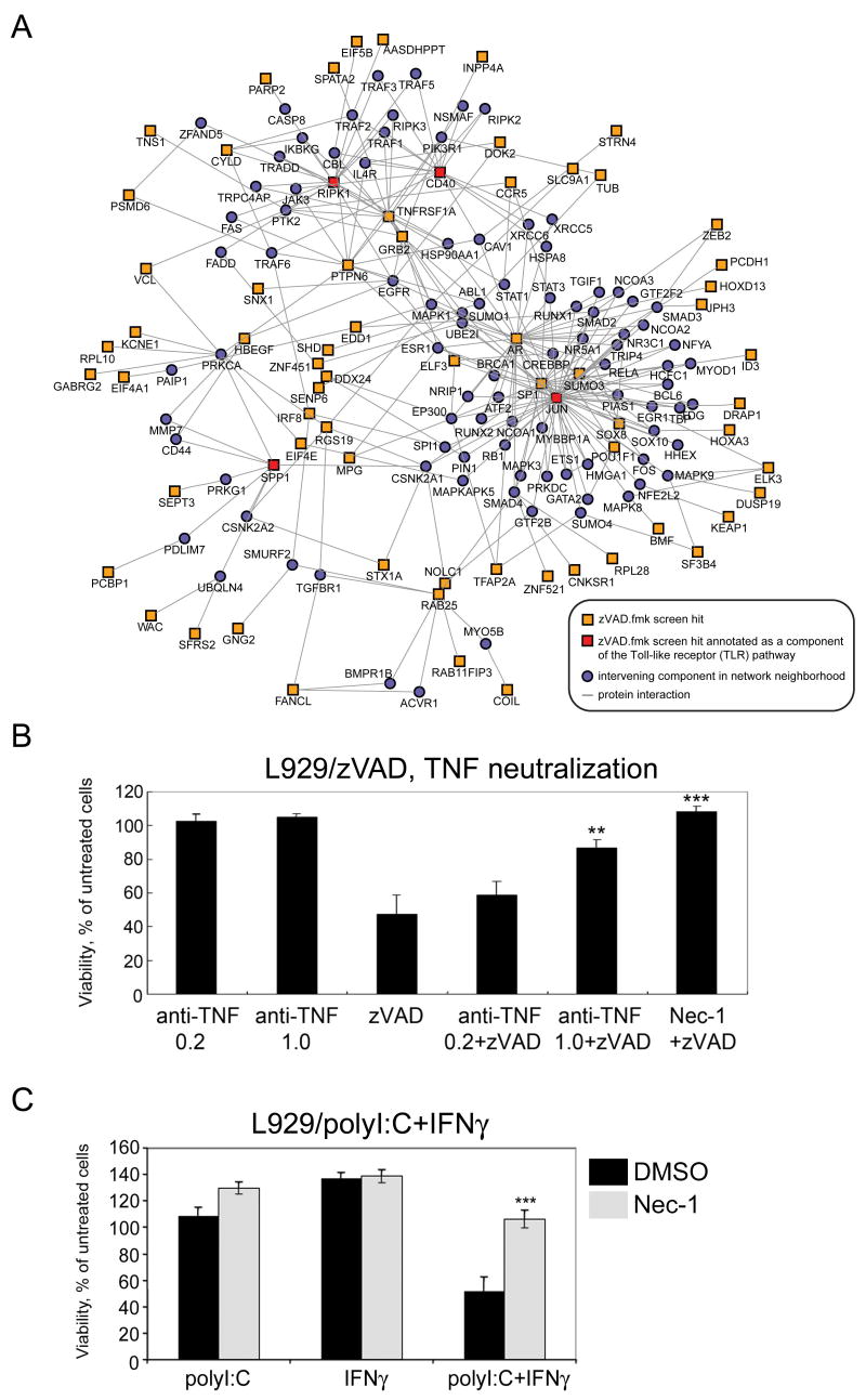
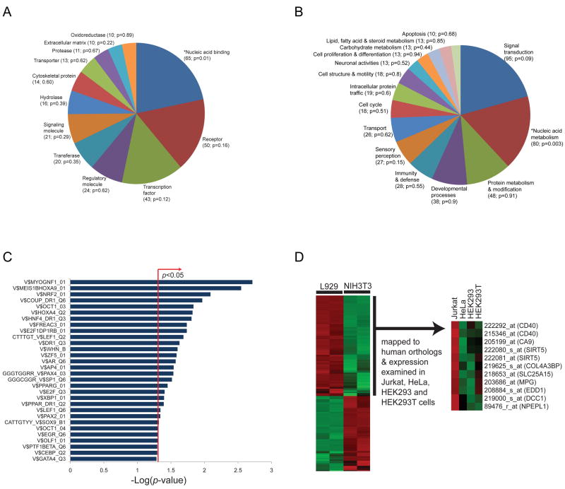
Stimulation of death receptors by agonists such as FasL and TNFalpha activates apoptotic cell death in apoptotic-competent conditions or a type of necrotic cell death dependent on RIP1 kinase, termed necroptosis, in apoptotic-deficient conditions. In a genome-wide siRNA screen for regulators of necroptosis, we identify a set of 432 genes that regulate necroptosis, a subset of 32 genes that act downstream and/or as regulators of RIP1 kinase, 32 genes required for death-receptor-mediated apoptosis, and 7 genes involved in both necroptosis and apoptosis. We show that the expression of subsets of the 432 genes is enriched in the immune and nervous systems, and cellular sensitivity to necroptosis is regulated by an extensive signaling network mediating innate immunity. Interestingly, Bmf, a BH3-only Bcl-2 family member, is required for death-receptor-induced necroptosis. Our study defines a cellular signaling network that regulates necroptosis and the molecular bifurcation that controls apoptosis and necroptosis.


Figures

Figure 1
Figure 1. siRNA screen for genes required for necroptosis
(A) A Schematic diagram of 1st and 2nd screens. (B) L929 cells, not-transfected (a), or transfected with either non-targeting siRNA(ctrl) or RIP1 siRNA at 50nM for 48hr (b), were treated with 20μM zVAD.fmk with or without 30μM Nec-1 for 18hrs. Lysates of siRNA transfected-L929 were analyzed by western blotting using anti-RIP1 or anti-Tubulin (bottom panel). (C) The morphological change of L929 cells transfected with indicated siRNAs after 18hr treatment of 20μM zVAD.fmk were shown by phase-contrast microscopic pictures. Scale bars: 100μm. Cellular viability was evaluated by measuring ATP levels in surviving cells using CellTiter-Glo. SD of cellular viability or cell death assays in all figures: *= p<0.05, **= p<0.01, ***= p<0.001, (n=4).
Figure 2
Figure 2. Expression patterns of the zVAD hits in mouse tissues and primary cells
(A) Expression profiles of genes from the zVAD.fmk screen showing significantly higher expression in immune cells or neuronal cells (FDR-adjusted p<0.05) as observed in the Novartis GNF1m microarray data examining expression across 61 mouse tissues. (B) Expression profiles of genes from the zVAD.fmk screen exhibiting increased expression in mouse macrophages, dendritic cells and NK cells or in B- and T-lymphocytes, from a large microarray panel of 119 mouse cell/tissue samples obtained from the RIKEN resource. (C) Involvement of necroptosis in primary macrophage cell death induced by zVAD.fmk. Isolated peritoneal macrophages from mice stimulated by thioglycollate were untreated or treated with 100μM zVAD.fmk with or without 30μM Nec-1 for 24hr and cell death was measured by Sytox assay (Invitrogen).
Figure 3
Figure 3. CYLD knockdown inhibits necroptosis
(A) L929 cells were transfected with 3 different siRNAs against cyld (cyld9 & cyld11), and rip1 (50nM) for 48hr, treated with 20μM zVAD.fmk for additional 18hr and the viability was measured as in Figure 1. Knockdown efficiency of cyld was confirmed by western blot using anti-CYLD, anti-RIP1 or anti-Tubulin antibodies. (B) Human FADD-deficient Jurkat cells were electroporated with siRNAs against human cyld (cyld6 & cyld7). Forty-eight hrs later, the cells were treated with 10ng/ml TNFα for additional 16hr and the viability was measured as in Figure 1. The cell lysates were analyzed by western blotting as in (A). (C) Inhibition of LC3-II induction in cyld-knockdown cells. L929 cells stably expressing shRNA for cyld(sh-cyld) or vector alone(vector) were treated with 20uM zVAD.fmk for indicated periods and the cell lysates were analyzed by western blotting using anti-LC3-II, anti-CYLD or anti-Tubulin antibodies.
Figure 4
Figure 4. Network extension of the innate immune Toll-like receptor (TLR) signaling pathway and its participation in necroptosis
(A) Network was constructed by anchoring on TLR pathway components (CD40, RIPK1, JUN and SPP1) that are also zVAD.fmk hits, using protein-protein interaction data curated from literature and high-throughput yeast-two-hybrid screens. (B) Autocrine production of TNFα is involved in zVAD-induced necroptosis. L929 cells were pretreated with indicated concentrations of anti-mouse TNFα antibody (μg/ml) for 1hr followed by treatment with or without 20μM zVAD.fmk or 30μM Nec-1 for 16hr. (C) Involvement of TLR pathway in necroptosis. L929 cells were treated with indicated chemicals for 19hr. Cellular viability was measured as described in Figure 1A. polyI:C; 25μg/ml, interferon gamma (IFNγ); 1000U/ml, Nec-1; 30μM.
Figure 5
Figure 5. Enrichment of transcription factor binding sites and nucleic acid binding function of genes involved in necroptosis
The 432 ‘hit’ genes from the secondary screen for zVAD.fmk-induced necroptosis were classified into (A) molecular function and (B) biological process categories for mouse genes according to the PANTHER classification system. Genes for which no annotations could be assigned were excluded from the analysis for both the hits and the global set (i.e. genes examined in the siRNA screen). Categories with at least 10 genes are displayed in the pie charts. The number of genes assigned to each category and enrichment p-values are shown in brackets. *denotes categories found enriched (unadjusted hypergeometric p<0.05) relative to the global set of genes examined in the screen. Lists of assigned genes grouped by molecular function or biological process categories are provided in Supplemental Tables 6 and 7, respectively. (C) Enrichment analysis of cis-regulatory elements, in particular transcription factor (TF) binding sites in the promoters of genes involved in zVAD.fmk-induced necroptosis (vertical axis of graph), using motif-based gene sets from the MSigDB and TF binding sites defined in the TRANSFAC database. TF-binding motifs were examined for enrichment among human orthologs of zVAD.fmk hits compared to the global set of genes screened in the siRNA library to which human orthologs could be mapped. The bar chart displays the negative log of the enrichment p-values for each pathway using the hypergeometric distribution. (D) Expression profiles of probes against zVAD.fmk screen hits showing elevated expression in necroptosis-sensitive L929 cells compared to necroptosis-resistant NIH3T3 cells were mapped to human orthologs and expression trends examined in four human cell lines: necroptosis-sensitive Jurkat cells, and necroptosis-resistant HeLa, HEK293 and HEK293T cells (from the GNF microarray collection). Expression values were z-score-transformed for each probe across arrays.
Figure 6
Figure 6. The Genes regulate necroptosis and apoptosis
(A) L929 cells were treated with 40ng/ml TNFα for 20hr with or without co-treatment of 30μM Nec-1. L929 cells, transfected with indicated siRNAs (tnfr; tnf receptor, ctrl; non-targeting siRNA), were treated with 10ng/ml TNFα for 18hr. Knockdown efficiency of TNFα receptor in L929 cells was determined by RT-PCR (right bottom panel). (B) Knockdown of Parp-2 inhibits necroptosis. L929 cells transfected with indicated siRNAs, treated with 20mM zVAD.fmk (left panel) for 18hr or with 10ng/ml TNFα for 20hr (right panel). The knockdown efficiency was determined by RT-PCR using Parp-1 as a control (left bottom panel). (C) Knockdown of Bmf inhibits necroptosis. L929 cells tranfected with indicated siRNAs (bmf, ctrl, cyld, rip1) were treated as in B. Knockdown efficiency of Bmf was determined by RT-PCR (left bottom panel) (D) Knockdown of Tipe1 inhibits necroptosis and apoptosis. L929 cells transfected with indicated siRNAs (tipe1, tnfr, cyld, rip1) were treated as in B. (E) NIH3T3 cell transfected with indicated siRNAs (tipe1, tnfr, rip1, cyld) (40nM) were treated with 10ng/ml TNFα+1.0μg/ml CHX for 12hr with or without co-treatment of 100μM zVAD,fmk (left bottom panel). Knockdown efficiency was determined by RT-PCR for Tipe1 using Tipe2 as a control (left upper and right bottom panels) or western blotting for RIP1, CYLD (left bottom panel). Cellular viability was measured as described in Figure1.

Comment in

References

    1. Cheng KW, Lahad JP, Kuo WL, Lapuk A, Yamada K, Auersperg N, Liu J, Smith-McCune K, Lu KH, Fishman D, et al. The RAB25 small GTPase determines aggressiveness of ovarian and breast cancers. Nat Med. 2004;10:1251–1256. - PubMed
    1. Clarke PG. Developmental cell death: morphological diversity and multiple mechanisms. Anat Embryol (Berl) 1990;181:195–213. - PubMed
    1. Degterev A, Hitomi J, Germscheid M, Ch'en IL, Korkina O, Teng X, Abbott D, Cuny GD, Yuan C, Wagner G, et al. Identification of RIP1 kinase as a specific cellular target of necrostatins. Nat Chem Biol. 2008;4:313–321. - PMC - PubMed
    1. Degterev A, Huang Z, Boyce M, Li Y, Jagtap P, Mizushima N, Cuny GD, Mitchison TJ, Moskowitz MA, Yuan J. Chemical inhibitor of nonapoptotic cell death with therapeutic potential for ischemic brain injury. Nat Chem Biol. 2005;1:112–119. - PubMed
    1. Fiers W, Beyaert R, Boone E, Cornelis S, Declercq W, Decoster E, Denecker G, Depuydt B, De Valck D, De Wilde G, et al. TNF-induced intracellular signaling leading to gene induction or to cytotoxicity by necrosis or by apoptosis. J Inflamm. 1995;47:67–75. - PubMed

Publication types

Substances