. 2009 Feb 6;385(5):1433-44.
doi: 10.1016/j.jmb.2008.12.017. Epub 2008 Dec 14.

Design and signaling mechanism of light-regulated histidine kinases

Affiliations

Design and signaling mechanism of light-regulated histidine kinases

Andreas Möglich et al. J Mol Biol. .

Abstract

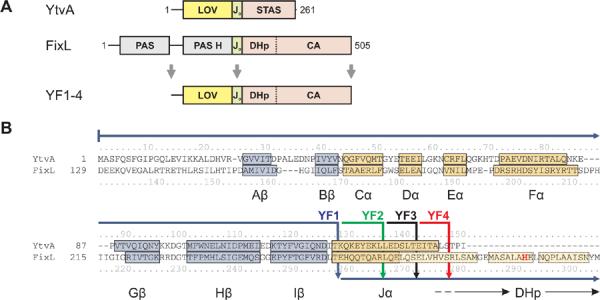
Signal transduction proteins are organized into sensor (input) domains that perceive a signal and, in response, regulate the biological activity of effector (output) domains. We reprogrammed the input signal specificity of a normally oxygen-sensitive, light-inert histidine kinase by replacing its chemosensor domain by a light-oxygen-voltage photosensor domain. Illumination of the resultant fusion kinase YF1 reduced net kinase activity by approximately 1000-fold in vitro. YF1 also controls gene expression in a light-dependent manner in vivo. Signals are transmitted from the light-oxygen-voltage sensor domain to the histidine kinase domain via a 40 degrees -60 degrees rotational movement within an alpha-helical coiled-coil linker; light is acting as a rotary switch. These signaling principles are broadly applicable to domains linked by alpha-helices and to chemo- and photosensors. Conserved sequence motifs guide the rational design of light-regulated variants of histidine kinases and other proteins.


Figures

Figure 1
Figure 1
Design of fusion proteins. A. YtvA comprises a LOV sensor and a STAS effector domain which are connected by the linker Jα. FixL consists of two PAS domains of which the second binds heme (PAS H) and a histidine kinase which comprises phosphoacceptor (DHp) and catalytic (CA) domains. Fusion kinases YF1-4 were obtained by linking the YtvA LOV domain to the FixL histidine kinase. B. Structure-based sequence alignment between YtvA and FixL. α-helices and β-strands within the LOV/PAS sensor domains are labelled and shown in yellow and blue; lighter shaded colors denote predictions from homology models. The active-site histidine 291 is highlighted in bold red. Solid arrows indicate how the two domains are fused in constructs YF1-4. An alignment spanning the full sequences of YtvA and FixL is given in Suppl. Fig. 1.
Figure 2
Figure 2
Kinase activity of fusion kinase YF1. A. In consecutive steps, YF1 autophosphorylates and then transfers the phosphoryl group to the response regulator FixJ. B. YF1 phosphorylates FixJ in the dark but is strongly inhibited in the light. Reactions were started by adding γ-32P-ATP and aliquots were taken at indicated times. Samples were separated on a SDS gel and incorporated phosphate monitored by 32P-radiography. Each lane contained 15 pmol YF1 and 380 pmol FixJ. C. Light absorption switches YF1 from kinase to phosphatase activity. Phosphorylation assays were started in the dark as in panel B (●). After 7.5 and 30.5 min aliquots of the reaction mix were exposed to light (dashed lines) which caused phospho-FixJ levels to decrease with initial velocities of (120 ± 20) (◯) and (140 ± 7) mol FixJ/(mol kinase·h) (▽). D. Comparison of reactions catalyzed by YF1 (●) and FixL (◯). Lines denote initial reaction velocities and correspond to turnovers of (56.4 ± 2.8) h−1 and (80.2 ± 3.6) h−1 for YF1 and FixL, respectively.
Figure 3
Figure 3
Kinase activity of YF1 depends on LOV photocycle. A. Upon illumination with 430 nm light (240 μW), the fraction of YF1 in the dark state as determined by absorption spectroscopy (◯) decays with a first-order time constant of (58.4 ± 1.0) s. YF1 kinase activity (●) decays with a time constant of (27.1 ± 1.0) s. B. Recovery kinetics of YF1 after saturating photobleaching. 70% of the absorption signal (thin line) recovers with a time constant of (5900 ± 25) s, followed by a slower phase. In contrast, kinase activity (●) displays sigmoid recovery kinetics which can be described by the model shown in panel C (thick line). C. Model for light inactivation of net kinase activity in dimeric YF1. D and L denote LOV domains in their dark and light states, respectively. Maximal kinase activity requires both YF1 LOV domains in the dark state. Photobleaching of one or both LOV domains strongly diminishes kinase activity. Analysis of YF1 recovery data according to this model yields a microscopic time constant τ of (10800 ± 1600) s.
Figure 4
Figure 4
A. Activity of fusion kinase variants in turnover assays. Blue bars denote activity in the dark and orange bars under constant illumination. Asterisks indicate activities below the detectable limit of 0.04 h−1 (dashed line). The table lists changes in helix angle and rise in the variants relative to YF1 in a canonical α-helix structure. Closely similar values are obtained when calculating these parameters based on a coiled coil model. B. Activity of linker variants as a function of helix angle relative to YF1. Variants with helix angles between −20° to +60° showed kinase activity in the dark; variants with angles between +40° to +100° were active in the light.
Figure 5
Figure 5
Multiple sequence alignment of PAS histidine kinases. A. Alignment of PAS histidine kinases that have 7n residues between the C-terminus of the PAS domain and the active-site histidine, indicated by the blue arrows. Out of 1811 sequences analyzed, 12 are shown and labeled with their UniProt identifiers. Residues conserved in more than half of all 1811 sequences are shown in bold red; brown shading denotes columns with more than 50% hydrophobic residues. The C-terminus of the PAS domains displays a highly conserved DIT consensus motif. The linker between the PAS and DHp domains shows the pattern of hydrophobic residues (labeled a and d) characteristic of α-helical coiled coils. Two gap positions have been arbitrarily inserted into the alignment to facilitate comparison with panel B. B. Alignment of PAS histidine kinases that have 7n+2 residues between the PAS domain and the active-site histidine. Out of 960 sequences analyzed, 12 are shown; B. japonicum FixL is in the bottom row (P23222). In comparison to panel A, the DHp domain differs in sequence and length around the phosphoacceptor histidine.

References

    1. Pawson T, Nash P. Assembly of cell regulatory systems through protein interaction domains. Science. 2003;300:445–52. - PubMed
    1. Dueber JE, Yeh BJ, Chak K, Lim WA. Reprogramming control of an allosteric signaling switch through modular recombination. Science. 2003;301:1904–8. - PubMed
    1. Bashor CJ, Helman NC, Yan S, Lim WA. Using engineered scaffold interactions to reshape MAP kinase pathway signaling dynamics. Science. 2008;319:1539–43. - PubMed
    1. Taylor BL, Zhulin IB. PAS domains: internal sensors of oxygen, redox potential, and light. Microbiol Mol Biol Rev. 1999;63:479–506. - PMC - PubMed
    1. Christie JM, Reymond P, Powell GK, Bernasconi P, Raibekas AA, Liscum E, Briggs WR. Arabidopsis NPH1: a flavoprotein with the properties of a photoreceptor for phototropism. Science. 1998;282:1698–701. - PubMed

Publication types

LinkOut - more resources