. 2009 Apr 1;45(2):614-26.
doi: 10.1016/j.neuroimage.2008.11.030. Epub 2008 Dec 9.

Functional connectivity of the human amygdala using resting state fMRI

Affiliations

Functional connectivity of the human amygdala using resting state fMRI

Amy Krain Roy et al. Neuroimage. .

Abstract

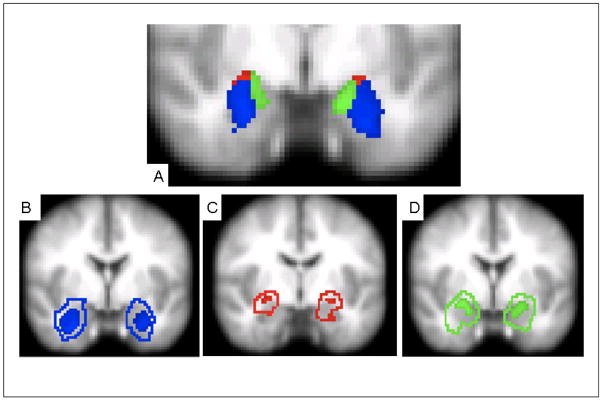
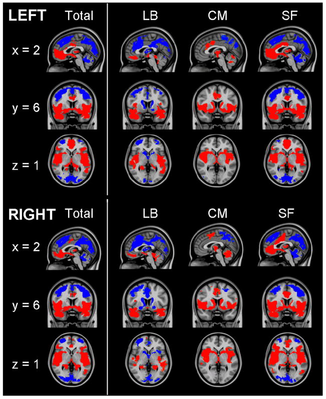
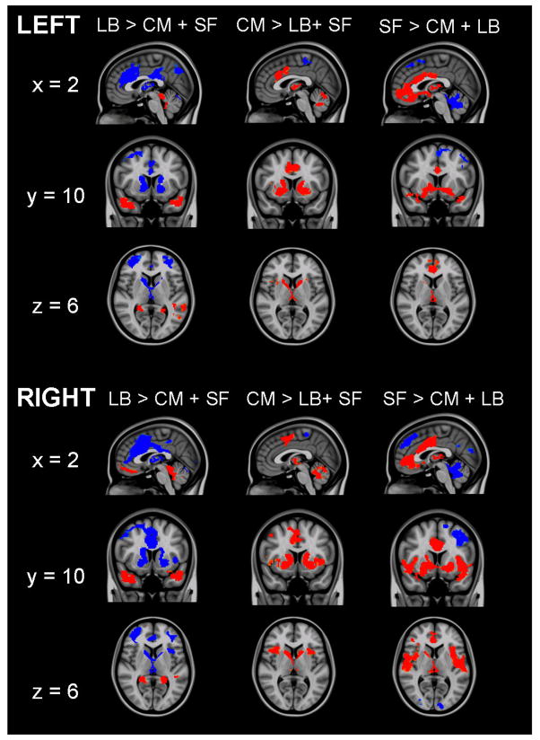
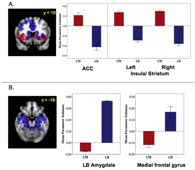
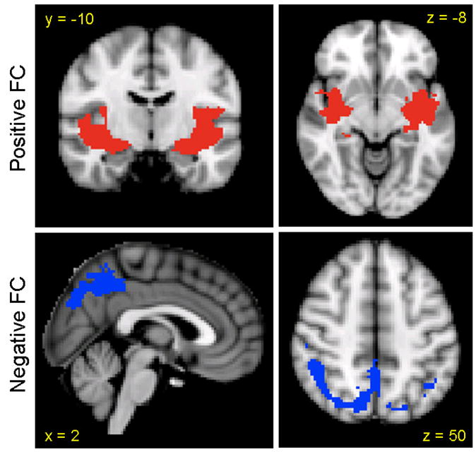
The amygdala is composed of structurally and functionally distinct nuclei that contribute to the processing of emotion through interactions with other subcortical and cortical structures. While these circuits have been studied extensively in animals, human neuroimaging investigations of amygdala-based networks have typically considered the amygdala as a single structure, which likely masks contributions of individual amygdala subdivisions. The present study uses resting state functional magnetic resonance imaging (fMRI) to test whether distinct functional connectivity patterns, like those observed in animal studies, can be detected across three amygdala subdivisions: laterobasal, centromedial, and superficial. In a sample of 65 healthy adults, voxelwise regression analyses demonstrated positively-predicted ventral and negatively-predicted dorsal networks associated with the total amygdala, consistent with previous animal and human studies. Investigation of individual amygdala subdivisions revealed distinct differences in connectivity patterns within the amygdala and throughout the brain. Spontaneous activity in the laterobasal subdivision predicted activity in temporal and frontal regions, while activity in the centromedial nuclei predicted activity primarily in striatum. Activity in the superficial subdivision positively predicted activity throughout the limbic lobe. These findings suggest that resting state fMRI can be used to investigate human amygdala networks at a greater level of detail than previously appreciated, allowing for the further advancement of translational models.


Figures

Figure 1
Figure 1
Location of 50% probabilistic masks of centromedial (CM), laterobasal (LB), and superficial (SF) amygdala projected on a mean EPI image using radiological convention. A) CM, LB, and SF masks (y = −6; Montreal Neurological Institute [MNI] standard space). B) 50% probabilistic mask of LB (solid) with extent of total LB according to the Juelich histological atlas as implemented in FSL (outline) (y = −2); C) 50% probabilistic mask of CM (solid) with extent of total CM according to the Juelich histological atlas as implemented in FSL (outline) (y = −8); D) 50% probabilistic mask of SF (solid) with extent of total SF according to the Juelich histological atlas as implemented in FSL (outline) (y = −2). Subdivision boundaries obtained from the Juelich histological atlas are based on probabilistic maps created by Amunts et al. (2005).
Figure 2
Figure 2
Functional connectivity of amygdala regions of interest. Patterns of significantly positive (red) and negative (blue) relationships for the total amygdala, laterobasal (LB), centromedial (CM) and superficial (SF) subdivisions. Sagittal (x = 2), coronal (y = 6), and axial (z = 1) views are presented. (MNI standard space; radiological convention; Z > 2.3; cluster significance: p < 0.05, corrected).
Figure 3
Figure 3
Direct comparisons of the functional connectivity of each subdivision with the other two subdivisions. Red indicates regions in which activity is significantly more positively predicted by spontaneous activity in target subdivision than by the other two subdivisions. Blue indicates regions in which activity is significantly more negatively predicted by spontaneous activity in the target subdivision than by the other two subdivisions. Sagittal (x = 2), coronal (y = 10), and axial (z = 6) views are presented. (MNI standard space; radiological convention; Z > 2.3; cluster significance: p < 0.05, corrected).
Figure 4
Figure 4
Hemispheric comparisons of functional connectivity. Mean parameter estimates represent the strength of FC between regions. (A) Hemispheric comparisons of the functional connectivity of the laterobasal subdivision of the amygdala with medial prefrontal cortex (PFC) and precentral gyrus. (B) Hemispheric comparisons of the functional connectivity of the centromedial subdivision of the amygdala with the SF and LB subdivisions, middle frontal gyrus, and medial frontal gyrus.
Figure 5
Figure 5
Regions showing opposing patterns of functional connectivity for LB and CM amygdala subdivisions in the right hemisphere (MNI standard space; radiological convention; Z > 2.3; cluster significance: p < 0.05, corrected). Mean parameter estimates represent the strength of FC between amygdala subdivision and region indicated. (A) Regions positively predicted by activity in the centromedial (CM) subdivision (red) and negatively predicted by activity in the laterobasal (LB) subdivision (blue) and their overlap (purple). ACC: anterior cingulate cortex. (B) Regions negatively predicted by activity in the centromedial (CM) subdivision (red) and positively predicted by activity in the laterobasal (LB) subdivision (blue) and their overlap (purple).
Figure 6
Figure 6
Regions of convergence among amygdala subdivisions. Regions of positive functional connectivity are indicated in red and regions of negative functional connectivity are indicated in blue (MNI standard space; radiological convention; Z > 2.3; cluster significance: p < 0.05, corrected).

References

    1. Achard S, Salvador R, Whitcher B, Suckling J, Bullmore E. A resilient, low-frequency, small-world human brain functional network with highly connected association cortical hubs. J Neurosci. 2006;26:63–72. - PMC - PubMed
    1. Akirav I, Maroun M. The role of the medial prefrontal cortex-amygdala circuit in stress effects on the extinction of fear. Neural Plast. 2007:30873. - PMC - PubMed
    1. Alexander GE, DeLong MR, Strick PL. Parallel organization of functionally segregated circuits linking basal ganglia and cortex. Annu Rev Neurosci. 1986;9:357–381. - PubMed
    1. Amaral DG. Amygdalohippocampal and amygdalocortical projections in the primate brain. Adv Exp Med Biol. 1986;203:3–17. - PubMed
    1. Amaral DG, Price JL. Amygdalo-cortical projections in the monkey (Macaca fascicularis) J Comp Neurol. 1984;230:465–496. - PubMed

Publication types