. 2009 Jan 30;33(2):215-26.
doi: 10.1016/j.molcel.2008.12.007. Epub 2008 Dec 24.

Cotranscriptional recruitment of the mRNA export factor Yra1 by direct interaction with the 3' end processing factor Pcf11

Affiliations

Cotranscriptional recruitment of the mRNA export factor Yra1 by direct interaction with the 3' end processing factor Pcf11

Sara Ann Johnson et al. Mol Cell. .

Abstract

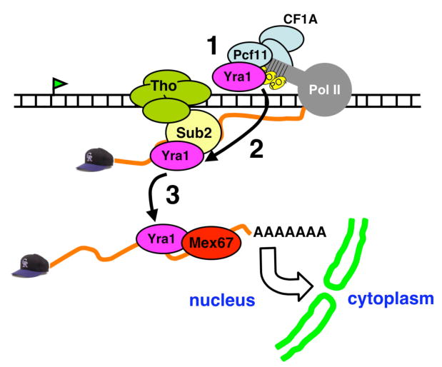
We investigated recruitment of the yeast mRNA export factor Yra1 to the transcription elongation complex (TEC). Previously, the Sub2 helicase subunit of TREX was proposed to recruit Yra1. We report that Sub2 is dispensable for Yra1 recruitment, but the cleavage/polyadenylation factor, CF1A, is required. Yra1 binds directly to the Zn finger/Clp1 region of Pcf11, the pol II CTD-binding subunit of CF1A, and this interaction is conserved between their human homologs. Tethering of Pcf11 to nascent mRNA is sufficient to enhance Yra1 recruitment. Interaction with Pcf11 can therefore explain Yra1 binding to the TEC independently of Sub2. We propose that after initially binding to Pcf11, Yra1 is transferred to Sub2. Consistent with this idea, Pcf11 binds the same regions of Yra1 that also contact Sub2, indicating a mutually exclusive interaction. These results suggest a mechanism for cotranscriptional assembly of the export competent mRNP and for coordinating export with 3' end processing.


Figures

Figure 1
Figure 1. Yra1 recruitment to actively transcribed genes is independent of Sub2
(A) ChIP analysis of WT, sub2-201 and sub2-206 cells at 37°C (90 minutes) with anti-pol II, anti-Sub2, anti-Yra1 and normal rabbit serum (NRS). 32P-labeled PCR products correspond to the indicated amplicons of TEF1. (B) Anti-Sub2 immunoblot (top panel) of WT and sub2 ts cells (10ug of whole cell extract (WCE)) at 25 and 37°C (90 minutes). Anti-Pgk1 immunoblot (bottom panel) serves as a loading control. Note that both Sub2 and Pgk1 protein levels increase slightly at 37°C in ALL cell types, including the WT. (C) ChIP analysis of PMA1 as in (A) analyzed by real-time PCR with amplicons shown in the map. Red lines on the map indicate putative poly A sites. Relative ChIP signals are plotted for Sub2 (top right), pol II (bottom left), and Yra1 (bottom right). Mean values, normalized to the maximal signal in each data set, +/- SEM are shown. (D) ChIP signals plotted as ratios of Yra1:pol II for PMA1and ACT1, +/- SEM.
Figure 2
Figure 2. Yra1 Recruitment is inhibited by inactivation of RNA15 and RNA14, and is not restored by deletion of the nuclear exosome
(A) ChIP analysis of WT rna15-2 and rna15-2Δrrp6 (DBY569) cells with anti-pol II, anti-Yra1 and NRS control. 32P-labeled PCR products of TEF1 amplicons as in Fig. 1A (see map) are shown. Asterisk (*) indicates non-specific product. Graph shows the ratio of Yra1:pol II ChIP signals determined by multiplex PCR, +/- SEM. (B) Anti-Yra1 immunoblot (top panel) of WT and mutant cells (10ug of WCE) at 25° and 37°C (60 minutes). The anti-Pgk1 immunoblot (bottom panel) serves as a loading control. (C) ChIP analysis of WT and rna14-1 and rna14-1Δrrp6 (DBY568) cells as in (A).
Figure 3
Figure 3. Co-transcriptional Recruitment of Yra1 Requires Functional Pcf11
(A) ChIP analysis of WT and pcf11-2 cells at 37° (60 minutes) with anti-pol II, anti-Sub2, anti-Yra1 and NRS control. 32P-labeled PCR products of TEF1 amplicons as in Fig. 1A (see map) are shown. In this experiment, after normalization to pol II, Yra1 occupancy at TEF1-5′ showed no change, TEF1-mid decreased 1.5 fold, and TEF1-3′ decreased 2.2 fold. (B) ChIP analysis of PMA1 as in Fig. 1C analyzed by real-time PCR with amplicons shown in the map. Red lines on the map indicate putative poly A sites. Relative ChIP signals are plotted for pol II (top left) and Yra1 (top right). Mean values, normalized to the maximal signal in each data set, +/- SEM are shown. (C) ChIP signals plotted as a ratios of Yra1:pol II and Sub2:pol II for PMA1, +/- SEM.
Figure 4
Figure 4. Yra1 Physically Interacts with CF1A via Pcf11
(A) Co-immunoprecipitation between Yra1 and CF1A from whole cell extracts (WCE). Antibodies used for IP are listed at the top, and those used for blotting are listed below each panel. IP's treated with RNase and DNase are indicated (+). IPs were washed with increasing concentrations of [Na+] (50mM, 100mM, 300mM, and 1000mM). Each sample represents the precipitation of 1mg WCE using 5ug antibody. (B) Yra1 binds specifically to the Pcf11 subunit of CF1A. GST pull-down assays using 35S-labeled in vitro translated Pcf11, RNA14, RNA15 and luciferase (Luc) negative control. Autoradiogram (top panel) and corresponding coomassie blue stained loading control (bottom panel) are shown. (C) Diagram of Pcf11 truncations used in (D) (see (Sadowski et al., 2003)). (D) Yra1 interacts with a C-terminal fragment of Pcf11 independent of the CID. GST pull-down analysis as in (B).
Figure 5
Figure 5. Conserved Binding of yeast and human Pcf11 to REF Family Proteins
(A) Alignment of yeast and human Pcf11 zinc finger/Clp1 regions. (B) Coomassie blue stained gel of His-tagged yeast and human Pcf11 fragments in (A). (C) Direct binding of yeast Pcf11 zinc finger/Clp1 region to Yra1. GST or GST-Yra1 bound to beads (see stained gel, bottom panel) was incubated with increasing amounts (1.25, 2.5, 5, 10ug) of yeast Pcf11(420-608), and bound protein was detected by anti-His immunoblot (top panel). Note the Pcf11(420-608) eluted from the beads migrates more slowly than the input (lane 1) because these samples contain 0.75M NaCl. (D). Direct binding of human Pcf11 zinc finger/Clp1 region to Aly. GST or GST-Aly bound to beads (see stained gel, bottom panel) was incubated with increasing amounts (1.25, 2.5, 5, 10ug) of human Pcf11(1342-1487), and bound protein was detected by anti-His immunoblot (top panel). (E) Domain diagram of full length Yra1 and truncations used in (F). The N/C REF protein fragment contains a linker (residues 92-132) from λ repressor. (F) Pull-down assays as in (C, D) with GST alone and fusions with full-length -Yra1, -N + RRM, -RRM, -RRM + C, -N, -C, and -N/C REF (see bottom panel). Yeast Pcf11(420-608) bound to the beads was eluted in high salt and visualized by anti-His immunoblot (top panel).
Figure 6
Figure 6. RNA tethering demonstrates that Yra1 and Pcf11 physically interact in vivo
(A) Diagram of Pcf11-HA-Npeptide tethering to nascent RNA via 6X BoxB sequences inserted in to the chromosomal GAL-YLR454W. (B) Tethering of Pcf11 to BoxB elements in GAL-YLR454W. ChIP analysis of non-tethered (YLR-6XBoxB/Pcf11:HA) and tethered (YLR-6XBoxB/Pcf11:HA-Npep) cells with anti-HA and NRS control. 32P-labeled PCR products corresponding to a region of GAL-YLR454W immediately adjacent to the tether (amplicon 1423) and to the 3′-end of the TEF1 gene are shown. (C) Pcf11 tethering alters Yra1 recruitment to the chromosomal GAL-YLR454W gene. Anti-Yra1 ChIP signals analyzed by real-time PCR for strains with tethered (purple bars) and non-tethered (blue bars) Pcf11. Diagram of GAL-YLR454W-6XBoxB and the corresponding amplicons are included below the plot. Data are represented as mean values, normalized to the maximal signal in the data set, +/- SEM. (D) Yra1 recruitment on a control gene PMA1 is unaffected by the Pcf11-HANpeptide tag. Plot of Yra1 occupancy quantified by real-time PCR and normalized to maximum values as in (C).
Figure 7
Figure 7. A revised model of co-transcriptional Yra1 recruitment by interaction with Pcf11 followed by hand-off to Sub2 and Mex67
1 = Recruitment of Yra1 via the Pcf11 subunit of CF1A. 2 = Transfer of Yra1 to the nascent RNA facilitated by binding of Sub2. 3 = Loading of the export receptor Mex67 onto the mRNA via interaction with Yra1.

Comment in

References

    1. Abruzzi KC, Belostotsky DA, Chekanova JA, Dower K, Rosbash M. 3′-end formation signals modulate the association of genes with the nuclear periphery as well as mRNP dot formation. EMBO J. 2006;25:4253–4262. - PMC - PubMed
    1. Abruzzi KC, Lacadie S, Rosbash M. Biochemical analysis of TREX complex recruitment to intronless and intron-containing yeast genes. EMBO J. 2004;23:2620–2631. - PMC - PubMed
    1. Ahn SH, Kim M, Buratowski S. Phosphorylation of serine 2 within the RNA polymerase II C-terminal domain couples transcription and 3′ end processing. Mol Cell. 2004;13:67–76. - PubMed
    1. Amrani N, Minet M, Wyers F, Dufour ME, Aggerbeck LP, Lacroute F. PCF11 encodes a third protein component of yeast cleavage and polyadenylation factor I. Mol Cell Biol. 1997;17:1102–1109. - PMC - PubMed
    1. Andrulis ED, Werner J, Nazarian A, Erdjument-Bromage H, Tempst P, Lis JT. The RNA processing exosome is linked to elongating RNA polymerase II in Drosophila. Nature. 2002;420:837–841. - PubMed

Publication types

MeSH terms

LinkOut - more resources