. 2009 Apr;57(4):373-82.
doi: 10.1369/jhc.2008.952895. Epub 2008 Dec 24.

Localization of mesenchymal cells in adult mouse thymus: their abnormal distribution in mice with disorganization of thymic medullary epithelium

Affiliations

Localization of mesenchymal cells in adult mouse thymus: their abnormal distribution in mice with disorganization of thymic medullary epithelium

Chikako Odaka. J Histochem Cytochem. 2009 Apr.

Abstract

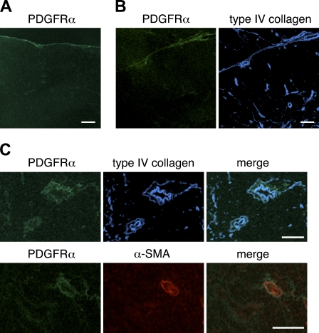
Thymic mesenchymal cells are known to be important for the development of the early fetal thymus into a functionally mature organ supporting T cell differentiation. We examined the expression of mesenchymal markers: pan-mesenchymal marker ER-TR7, desmin, alpha-smooth muscle actin (alpha-SMA), and alpha- and beta-chain of platelet-derived growth factor receptor (PDGFRalpha, PDGFRbeta) in thymi of normal adult mice. Desmin and ER-TR7 revealed specific staining in the capsule, septa, and perivascular cells. Most perivascular cells highly expressed PDGFRbeta at the same levels as desmin. Low expression of PDGFRalpha was detected in the capsule, intralobular septa, and some perivascular cells of normal adult thymi. alpha-SMA, used to identify vascular smooth muscle cells, was detectable on arterioles and some large venules but not on capillaries. Thus, desmin, PDGFRalpha, and PDGFRbeta were localized in the capsule, septa, and perivascular cells in thymus of adult mouse, although there were differences in the expression level among these markers. On the other hand, the expression of mesenchymal markers was detectable in the region of the thymic medullary epithelium of lymphotoxin beta receptor-deficient mice and plt/plt mice, indicating that mesenchymal cells were abnormally localized in the region. These results suggest that disorganization of the medullary epithelium may be accompanied by aberrant distribution of mesenchyme in adult mouse thymus.


Figures

Figure 1
Figure 1
Expression of desmin in the thymus of adult mouse. Immunofluorescence staining of thymic sections of adult BALB/c mice was performed to detect ER-TR7 (A) (green), CD31 (B) (green), desmin (A,B) (red), or type IV collagen (B) (blue). (A) Double staining for ER-TR7 fibroblast marker and desmin shows their colocalization along capsule and septa and around vasculature. (B) Desmin is detectable around CD31-positive blood vessels, and desmin-positive perivascular cells are associated with basement membranes. Bar = 100 μm.
Figure 2
Figure 2
Expression of α-smooth muscle actin (α-SMA) in the thymus of adult mouse. Immunofluorescence staining of thymic sections of adult BALB/c mice was performed to detect α-SMA (A,B) (green), CD31 (A,B) (red), or ephrinB2 (B) (blue). α-SMA reactivity is detectable on eprinB2-positive arterioles and some large ephrinB2-negative venules. α-SMA–positive vascular smooth muscle cells (VSMCs) on arterioles are uniformly shaped, closely packed, and tightly associated with the endothelium. Bar = 100 μm.
Figure 3
Figure 3
Expression of the α-chain of platelet-derived growth factor receptor (PDGFRα) in the thymus of adult mouse. Immunofluorescence staining of thymic sections of adult BALB/c mice was performed to detect PDGFRα (A,B,C) (green), α-SMA (B) (red), or type IV collagen (B,C) (blue). PDGFRα reactivity is weak but detectable in the capsule (A), along intralobular septa (B), and on perivascular cells including α-SMA–positive VSMCs (C). Bar = 100 μm.
Figure 4
Figure 4
Expression of PDGFRβ in the thymus of adult mouse. Immunofluorescence staining of thymic sections of adult BALB/c thymi was performed to detect PDGFRβ (A,B,C) (green), desmin (C) (red), or type IV collagen (B,C) (blue). PDGFRβ-positive cells are detectable in the capsule (A) and intralobular septa (B). PDGFRβ-positive cells are seen around almost all blood vessels, and most desmin-positive perivascular cells highly express PDGFRβ (C). Bar = 100 μm.
Figure 5
Figure 5
Organization of thymic medullary epithelium in adult C57BL/6 wild-type, lymphotoxin β receptor–negative (LTβR−/−), and plt/plt mice. Immunofluorescence staining of thymic sections of adult C57BL/6 wild-type, LTβR−/−, and plt/plt mice was performed to detect keratin 5 (green) and the binding to Ulex europaeus agglutinin-1 (UEA-1) (red). Aberrant medulla formation and marked reduction in UEA-1 binding to medullary epithelial cells are seen in LTβR−/− mice. Plt/plt thymi tend to have small medullary islets, but the clusters of medullary epithelial cells expressing keratin 5 and binding to UEA-1 are present. Bar = 100 μm.
Figure 6
Figure 6
Localization of ER-TR7 and medullary epithelium in the thymi of adult C57BL/6 wild-type, LTβR−/−, and plt/plt mice. (A,B) Immunofluorescence staining of thymic sections of adult C57BL/6 wild-type, LTβR−/−, and plt/plt mice was performed to detect ER-TR7 (green) and keratin 5 (red). Aberrant foci in labeling with ER-TR7 are evident in the thymic medullary region of both LTβR−/− and plt/plt mice. (B) High magnification. Many ER-TR7–positive cells are situated in epithelial cell–free regions of the thymic medulla and are found to be frequently adjacent to the epithelial cells, but coexpression of ER-TR7 and keratin 5 is rarely detectable in LTβR−/− thymi. Bar = 100 μm.
Figure 7
Figure 7
Localization of desmin, PDGFRβ, and medullary epithelium in the thymi of adult LTβR−/− mice. Immunofluorescence staining of thymic sections of adult LTβR−/− mice was performed to detect desmin (A) (green), or PDGFRβ (B) (green), and keratin 5 (A,B) (red). Many desmin- or PDGFRβ-expressing cells are situated in epithelial cell–free regions of thymic medulla. Bar = 100 μm

References

    1. Alpdogan O, Hubbard VM, Smith OM, Patel N, Lu S, Goldberg GL, Gray DH, et al. (2006) Keratinocyte growth factor (KGF) is required for postnatal thymic regeneration. Blood 107:2453–2460 - PMC - PubMed
    1. Bergers G, Song S (2005) The role of pericytes in blood-vessel formation and maintenance. Neuro Oncol 7:452–464 - PMC - PubMed
    1. Bockman DE, Kirby ML (1984) Dependence of thymus development on derivatives of the neural crest. Science 223:498–500 - PubMed
    1. Boehm T, Scheu S, Pfeffer K, Bleul CC (2003) Thymic medullary epithelial cell differentiation, thymocyte emigration, and the control of autoimmunity require lympho-epithelial cross talk via LTßR. J Exp Med 198:757–769 - PMC - PubMed
    1. Bondjers C, He L, Takemoto M, Norlin J, Asker N, Hellström M, Lindahl P, et al. (2006) Microarray analysis of blood microvessels from PDGF-B and PDGF-Rbeta mutant mice identifies novel markers for brain pericytes. FASEB J 20:1703–1705 - PubMed

MeSH terms