. 2009 Apr;57(4):383-96.
doi: 10.1369/jhc.2008.952853. Epub 2008 Dec 24.

Expression of Muc19/Smgc gene products during murine sublingual gland development: cytodifferentiation and maturation of salivary mucous cells

Affiliations

Expression of Muc19/Smgc gene products during murine sublingual gland development: cytodifferentiation and maturation of salivary mucous cells

Biswadip Das et al. J Histochem Cytochem. 2009 Apr.

Abstract

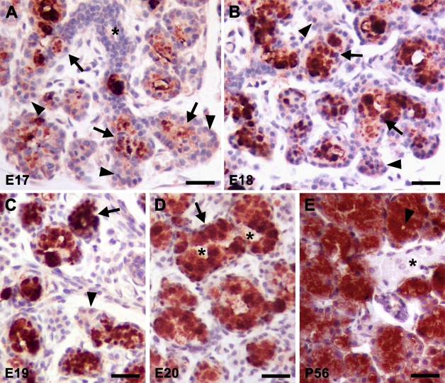
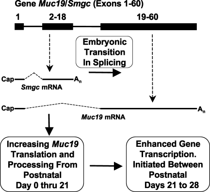
Muc19/Smgc expresses two splice variants, Smgc (submandibular gland protein C) and Muc19 (mucin 19), the latter a major exocrine product of differentiated murine sublingual mucous cells. Transcripts for Smgc were detected recently in neonatal sublingual glands, suggesting that SMGC proteins are expressed during initial salivary mucous cell cytodifferentiation. We therefore compared developmental expression of transcripts and translation products of Smgc and Muc19 in sublingual glands. We find abundant expression of SMGC within the initial terminal bulbs, with a subsequent decrease as Muc19 expression increases. During postnatal gland expansion, SMGC is found in presumptive newly formed acinar cells and then persists in putative acinar stem cells. Mucin levels increase 7-fold during the first 3 weeks of life, with little change in transcript levels, whereas between postnatal days 21 and 28, there is a 3-fold increase in Muc19 mRNA and heteronuclear RNA. Our collective results demonstrate the direct transition from SMGC to Muc19 expression during early mucous cell cytodifferentiation and further indicate developmentally regulated changes in Muc19/Smgc transcription, alternative splicing, and translation. These changes in Muc19/Smgc gene expression delineate multiple stages of salivary mucous cell cytodifferentiation and subsequent maturation during embryonic gland development through the first 4 weeks of postnatal life.


Figures

Figure 1
Figure 1
Expression of Muc19/Smgc gene products and gland mass during sublingual gland development. cDNA from each preparation of glands was assayed by real-time RT-PCR for submandibular gland protein C (Smgc) mRNA (A), mucin 19 (Muc19) mRNA (B), and Muc19 heteronuclear RNA (C). Transcript copy numbers/ng cDNA were normalized to 18S RNA and are shown as means ± SE. 18S RNA levels were consistent from embryonic day 17 (E17) and later (mean ± SE, 13.1 ± 0.2 × 10¹⁰ transcripts/ng cDNA). Values were lower at E15 (3.8 ± 1.1 × 10¹⁰ transcripts/ng cDNA) and E16 (6.0 ± 0.2 × 10¹⁰ transcripts/ng cDNA). (D) Gland wet weights. Values are means ± SE. Hatched bars, unsexed prenatal glands. Black bars, male glands. Open bars, female glands. Numbers over bars indicate gland preparations per age group (1 to 20 glands/preparation), with equal preparations for each sex. The same preparations were used in panels A–C, whereas additional preparations were incorporated for determining gland weights.
Figure 2
Figure 2
Immunohistochemical localization of SMGC during sublingual gland development. Ages are indicated in the lower left corner of each panel. (A) Staining in early terminal epithelial bulbs. Arrowheads, immunoreactive pyramidal cells at distal ends of branching tubuloacini. Arrow, cells in more-proximal regions, with basal nuclei and larger cytoplasm that display intermediate staining. Asterisk, duct containing a central lumen within branching tubuloacinus. (B) A day later, proximal cells of terminal bulbs are more abundant and display moderate flocculent-like cytoplasmic staining (arrows). Arrowhead, strong immunoreactivity by small pyramidal cells at the distal end of a branching tubuloacinus. (C) Staining is more variable at E19. Arrowheads, acinar cells with intense staining. Arrow, acinus with lighter cytoplasmic staining. (D) Mucous tubuloacini are apparent shortly before birth. Asterisk, a branched V-shaped mucous tubuloacinus with light granular cytoplasmic staining. Arrow, fully mature mucous cell with an unstained and expanded cytoplasm. (E) Light staining in the perinuclear region of mucous acinar cells at 3 days of age. Arrowhead, acinar cell with strong granular cytoplasmic staining. (F) Mucous acinar cells are mostly unstained. Arrowheads, rare example of a group of strongly stained acinar cells. Arrow, two adjoining intercalated ducts in cross section. (G) Most cells are unstained at 4 weeks of age. Arrowhead, light perinuclear staining of an acinar cell. Arrow, subpopulation of intercalated duct cells with light staining. Asterisk, lumen of intercalated duct. (H) Strong staining of a subpopulation of cells lining the lumen of a large embryonic extralobular excretory duct. See Results for details. Vector Red substrate with hematoxylin counterstain in all cases. Bar = 35 μm.
Figure 3
Figure 3
Expression of transcripts for large mucins in adult and neonatal (3-day-old) sublingual glands. RT-PCR was performed using 50 ng of cDNA, 30 cycles, and primers specific for the indicated mucin transcripts (see Materials and Methods for details). Shown are representative results from three independent preparations of cDNA from each age group. Tissues serving as positive controls for transcripts absent in sublingual glands were ileum (Muc2), stomach (Muc5ac and Muc6), tracheolarynx (Muc5b), and ovaries (Muc16).
Figure 4
Figure 4
Immunohistochemical localization of Muc19 during sublingual gland development. Ages are indicated in the lower left corner of each panel. (A) Staining in early terminal bulbs is localized to the apical cytoplasm of the larger proximal cells (arrows). Arrowheads, absence of staining in pyramidal cells at distal ends of branching tubuloacini. Asterisk, lumen of duct at proximal end of terminal tubule. (B) Immunoreactive proximal cells are more abundant at E18 (arrows). Distal ends of terminal bulbs are unstained (arrowheads). (C) Acini with strongly stained columnar cells are apparent the following day (arrow). Arrowhead, unstained cells at the distal end of a terminal bulb. (D) Mucous tubuloacini are apparent at E20 and display strong immunoreactivity. Two asterisks demark the ends of a lumen within a large mucous tubuloacinus. Arrow, tall columnar mucous acinar cells with basally compressed nuclei and strongly stained cytoplasm. (E) Intensely stained mucous tubuloacini of an adult gland. Arrowhead, acinar lumen. Asterisk, unstained ductal structure. See Results for details. Vector Red substrate with hematoxylin counterstain in all cases. Bar = 35 μm.
Figure 5
Figure 5
Western blot of SMGC in whole-gland homogenates at postnatal days 3 through 56. Postnatal days of age and sexes are indicated above the lanes. Far left lane is a control (Con) from a 3-day-old submandibular gland. All lanes were loaded with the equivalent of 300 μg wet weight of tissue and run on a 4–12% gradient gel. The blot was initially exposed to film for 30 sec. The lanes corresponding to ages 28 days and 56 days were re-exposed to film for 4 min to distinguish less-abundant proteins. Mobilities of molecular mass markers (kDa) are indicated on the left.
Figure 6
Figure 6
Muc19 in whole-gland homogenates at postnatal days 3 through 56. Three separate sample preparations from each age group were run on 4–12% SDS-PAGE gels (50 μg wet weight of tissue/lane) and stained with Alcian blue followed by silver enhancement. (A) Image of the stained glycoproteins in each lane. Ages are given above the lanes. (B) Graph of the net mean pixel intensities in the lanes shown in A, expressed as mean ± SE for the triplicate values of each age group.
Figure 7
Figure 7
Evaluation of t-Smgc transcripts in 8-week-old mice. (A) Semi-quantitative comparison of Muc19 and t-Smgc transcripts in sublingual glands. Increasing amounts of random primed cDNA (x-axis) were probed by PCR (35 cycles) with primers to Smgc (exons 1 and 18) and the 3′ end of Muc19. Products were run on a 1.5% agarose gel (insert). Net mean pixel intensity of each band was determined and plotted versus the amount of input cDNA. Slopes of the lines (net mean pixel intensity/ng cDNA) are from linear regressions (r2 > 0.98) and were 4.0 × 10⁵ (Muc19) and 1.8 × 10² (t-Smgc). (B) Sublingual gland t-Smgc transcripts are associated with poly(A+) RNA. Double-selected poly(A+) and poly(A) RNA fractions and total RNA were added to reverse transcription reactions in the presence (+) and absence (−) of reverse transcriptase, then products were subjected to PCR with primers specific for Smgc. PCRs specific for Muc19 and 18S RNA serve as positive controls for poly(A+) and poly(A) RNA selective transcripts, respectively. Results are representative of two independent experiments. (C) Transcripts for t-Smgc are not selectively localized to nuclei. Sublingual glands were fractionated into nuclei-containing and nuclei-free fractions. Random primed cDNA prepared from each fraction was PCR amplified with primers specific for t-Smgc. Results of three independent preparations are shown. (D) Transcripts for t-Smgc (823 bp product, upper panel) and Muc19 are readily detectable by PCR (35 cycles) in random primed cDNA (10 ng) from adult sublingual glands (SLG) but not in submandibular (SMG) or parotid glands. Left side of t-Smgc panel, mobilities of 1 kb ladder. Bottom panel, β-actin controls. See Materials and Methods for details.
Figure 8
Figure 8
Working model of key mechanisms controlling differential Muc19/Smgc gene expression in salivary mucous cells during cytodifferentiation and maturation. A simplified diagram of the gene (top) indicates exon 1 (shared by Smgc and Muc19 transcripts) and two boxes representing exons exclusively utilized either for Smgc (2–18) or for Muc19 (19–60) transcripts. Not drawn to scale. See text for details.

References

    1. Ball WD, Hand AR, Johnson AO (1988a) Secretory proteins as markers for cellular phenotypes in rat salivary glands. Dev Biol 125:265–279 - PubMed
    1. Ball WD, Hand AR, Moreira JE, Johnson AO (1988b) A secretory protein restricted to type I cells in neonatal rat submandibular glands. Dev Biol 129:464–475 - PubMed
    1. Ball WD, Redman RS (1984) Two independently regulated secretory systems within the acini of the submandibular gland of the perinatal rat. Eur J Cell Biol 33:112–122 - PubMed
    1. Chen Y, Zhao YH, Kalaslavadi TB, Hamati E, Nehrke K, Le AD, Ann DK, et al. (2004) Genome-wide search and identification of a novel gel-forming mucin MUC19/Muc19 in glandular tissues. Am J Respir Cell Mol Biol 30:155–165 - PubMed
    1. Culp DJ, Latchney LR, Fallon M, Denny PA, Couwenhoven PC, Sally RI, Chuang S (2004) The gene encoding mouse Muc19: cDNA, genomic organization and relationship to Smgc. Physiol Genomics 19:303–318 - PubMed

Publication types