. 2009 Jan;49(1):287-96.
doi: 10.1002/hep.22678.

The pathogen receptor liver and lymph node sinusoidal endotelial cell C-type lectin is expressed in human Kupffer cells and regulated by PU.1

Affiliations

The pathogen receptor liver and lymph node sinusoidal endotelial cell C-type lectin is expressed in human Kupffer cells and regulated by PU.1

Angeles Domínguez-Soto et al. Hepatology. 2009 Jan.

Abstract

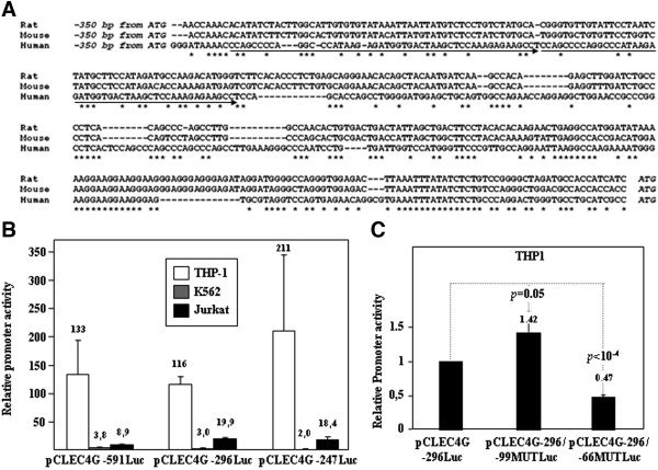
Human LSECtin (liver and lymph node sinusoidal endothelial cell C-type lectin, CLEC4G) is a C-type lectin encoded within the L-SIGN/DC-SIGN/CD23 gene cluster. LSECtin acts as a pathogen attachment factor for Ebolavirus and the SARS coronavirus, and its expression can be induced by interleukin-4 on monocytes and macrophages. Although reported as a liver and lymph node sinusoidal endothelial cell-specific molecule, LSECtin could be detected in the MUTZ-3 dendritic-like cell line at the messenger RNA (mRNA) and protein level, and immunohistochemistry analysis on human liver revealed its presence in Kupffer cells coexpressing the myeloid marker CD68. The expression of LSECtin in myeloid cells was further corroborated through the analysis of the proximal regulatory region of the human LSECtin gene, whose activity was maximal in LSECtin+ myeloid cells, and which contains a highly conserved PU.1-binding site. PU.1 transactivated the LSECtin regulatory region in collaboration with hematopoietic-restricted transcription factors (Myb, RUNX3), and was found to bind constitutively to the LSECtin proximal promoter. Moreover, knockdown of PU.1 through the use of small interfering RNA led to a decrease in LSECtin mRNA levels in THP-1 and monocyte-derived dendritic cells, thus confirming the involvement of PU.1 in the myeloid expression of the lectin.

Conclusion: LSECtin is expressed by liver myeloid cells, and its expression is dependent on the PU.1 transcription factor.


Figures

Figure 1
Figure 1
Expression of LSECtin in the MUTZ‐3 cell line. (A) Detection of LSECtin, DC‐SIGN, and GAPDH mRNA in untreated and IL‐4–treated MUTZ‐3 via conventional RT‐PCR. The results of control experiments without RNA (H2O) or without reverse‐transcriptase (CNT RT) are shown in each case. (B) Relative levels of LSECtin mRNA in untreated (−) and IL‐4–treated MUTZ‐3 cells via quantitative RT‐PCR, after normalization for the levels of 18S RNA. Determination was performed in triplicate, and the mean and standard deviation is shown. (C) Detection of DC‐SIGN and LSECtin protein expression in untreated (−) and IL‐4–treated MUTZ‐3 cells via western blotting, using a polyclonal antiserum against their corresponding neck regions. For control purposes, the expression of both lectins in MDDCs and untransfected or LSECtin‐transfected COS‐7 is shown.
Figure 2
Figure 2
LSECtin expression in human liver. Immunolocalization of LSECtin, PU.1, CD68, and CD31 on formalin‐fixed, paraffin‐embedded human liver tissue sections. LSECtin was detected with rabbit polyclonal antisera against its extracellular region (ADS4). The upper right panel shows the staining yielded by a preimmune rabbit antiserum. Arrowheads indicate the position of LSECtin‐ or CD68‐positive cells in their respective panels.
Figure 3
Figure 3
LSECtin expression in human lymph nodes. (A) Immunolocalization of LSECtin, factor XIII (FXIII) CD68, CD31, and factor VIII (FVIII) on formalin‐fixed, paraffin‐embedded human lymph node tissue sections (magnification ×20). LSECtin was detected with rabbit polyclonal antisera against the neck domain (ADS1, upper panels). (B,C) Detection of LSECtin‐positive cells in human lymph node tissue section areas enriched in CD68+ cells. (C) The areas marked by boxes are shown at a higher magnification in the lower panels (magnification ×40).
Figure 4
Figure 4
Coexpression of LSECtin and the CD68 myeloid‐specific marker in human liver tissue sections. (A) Simultaneous immunolocalization of LSECtin (red) and CD68 (brown) on formalin‐fixed, paraffin‐embedded human liver tissue sections (magnification ×40). LSECtin was detected with rabbit polyclonal antisera against the extracellular region (ADS4), and CD68 was detected with the PG‐M1 monoclonal antibody. Two different preparations are shown (middle panels). The areas marked by boxes are shown in the lower panels at higher magnification (magnification ×100). The staining yielded by each individual antibody in the presence of the corresponding negative controls is shown in the upper panels. (B) Relative levels of PU.1, CD68, and LSECtin mRNA in human hepatocytes, sinusoidal endothelial cells (HSEC), Kupffer cells, and Ito cells via quantitative RT‐PCR after normalization for the levels of GAPDH RNA. Determination was performed in triplicate, and the mean and standard deviation is shown.
Figure 5
Figure 5
Structural and functional analysis of the LSECtin gene proximal regulatory region. (A) Sequence alignment of the proximal regulatory regions (from −350 to the translation initiation site) of the human, murine, and rat LSECtin genes. Identities are shown by asterisks below the sequences. The position of the 48‐nucleotide direct repeats within the human LSECtin promoter is indicated by arrows below the sequence. (B) THP‐1, K562, and Jurkat cells were transfected with the indicated reporter plasmids, and luciferase activity was determined after 24 hours. Promoter activity is expressed relative to the activity produced by the promoterless pXP2 plasmid in each cell type and after normalization for transfection efficiency. Data represent the mean ± SD of four independent experiments using two different DNA preparations. (C) THP‐1 cells were transfected with the indicated reporter plasmids, and luciferase activity was determined after 24 hours. Promoter activity is expressed relative to the activity produced by the wild‐type pCLEC4G −296Luc construct after normalization for transfection efficiency. Data represent the mean ± SD of four independent experiments.
Figure 6
Figure 6
PU.1 binds in vivo and modulates the activity of the LSECtin promoter in vitro. (A,B) NIH‐3T3 cells were transfected with the indicated LSECtin promoter‐based reporter plasmids in the presence of an empty expression vector (pCDNA3.1) or expression vectors for RUNX3, Myb, or PU.1, either alone or in combination. For each individual reporter construct, fold induction represents the luciferase activity yielded by each expression vector combination relative to the activity produced by an identical amount of empty CMV‐0 plasmid. In all cases, total DNA was kept constant (1.5 μg) by adding CMV‐0 plasmid DNA, and luciferase activity was determined after 24 hours. Data represent the mean ± standard deviation of three independent experiments using two different DNA preparations. (C) In vivo occupancy of the LSECtin proximal promoter by PU.1. Shown are chromatin immunoprecipitations on immature monocyte‐derived DCs using an affinity‐purified polyclonal antiserum specific for PU.1, a nonspecific affinity‐purified antiserum (control Ab), or no antibody (no Ab). Immunoprecipitated chromatin was analyzed via PCR using a pair of LSECtin promoter‐specific primers that together amplify 312‐bp and 263‐bp fragments spanning from −296/−247 to +16, since the forward primer anneals to a 48‐nucleotide direct repeat. Input DNA lanes represent the PCR analysis performed on DNA from a 1:20 dilution of the starting sonicated lysate. Additional controls include amplification in the absence of DNA (no DNA) or amplification of human genomic DNA (genomic DNA).
Figure 7
Figure 7
Knockdown on PU.1 results in diminished LSECtin mRNA levels. (A) THP‐1 cells or (B) MDDCs were nucleofected with either siRNA for PU.1 (siRNA PU.1) or a control siRNA (siRNA CNT). After 24 hours, total RNA was isolated and LSECtin mRNA was measured via quantitative RT‐PCR. Each experiment was performed in duplicate, and both experiments are shown. Results are expressed as the relative LSECtin mRNA level, which indicates the level of LSECtin mRNA in each sample relative to its level in control siRNA‐nucleofected cells. To confirm siRNA efficiency in each individual experiment, one‐third of the cells were lysed and underwent western blotting (inserts), using a polyclonal antiserum against human PU.1 and a β‐actin–specific monoclonal antibody for loading control purposes.

References

    1. Geijtenbeek TB, Torensma R, van Vliet SJ, van Duijnhoven GC, Adema GJ, van Kooyk Y, et al. Identification of DC‐SIGN, a novel dendritic cell‐specific ICAM‐3 receptor that supports primary immune responses. Cell 2000; 100: 575–585. - PubMed
    1. Soilleux EJ, Barten R, Trowsdale J. DC‐SIGN; a related gene, DC‐SIGNR; and CD23 form a cluster on 19p13. J Immunol 2000; 165: 2937–2942. - PubMed
    1. Bashirova AA, Geijtenbeek TB, van Duijnhoven GC, van Vliet SJ, Eilering JB, Martin MP, et al. A dendritic cell‐specific intercellular adhesion molecule 3‐grabbing nonintegrin (DC‐SIGN)‐related protein is highly expressed on human liver sinusoidal endothelial cells and promotes HIV‐1 infection. J Exp Med 2001; 193: 671–678. - PMC - PubMed
    1. Liu W, Tang L, Zhang G, Wei H, Cui Y, Guo L, et al. Characterization of a novel C‐type lectin‐like gene, LSECtin: demonstration of carbohydrate binding and expression in sinusoidal endothelial cells of liver and lymph node. J Biol Chem 2004; 279: 18748–18758. - PubMed
    1. Dominguez‐Soto A, Aragoneses‐Fenoll L, Martin‐Gayo E, Martinez‐Prats L, Colmenares M, Naranjo‐Gomez M, et al. The DC‐SIGN‐related lectin LSECtin mediates antigen capture and pathogen binding by human myeloid cells. Blood 2007; 109: 5337–5345. - PubMed

Publication types