. 2009 Mar;216(1):207-18.
doi: 10.1016/j.expneurol.2008.11.019. Epub 2008 Dec 10.

Neuritic dystrophy and neuronopathy in Akita (Ins2(Akita)) diabetic mouse sympathetic ganglia

Affiliations

Neuritic dystrophy and neuronopathy in Akita (Ins2(Akita)) diabetic mouse sympathetic ganglia

Robert E Schmidt et al. Exp Neurol. 2009 Mar.

Abstract

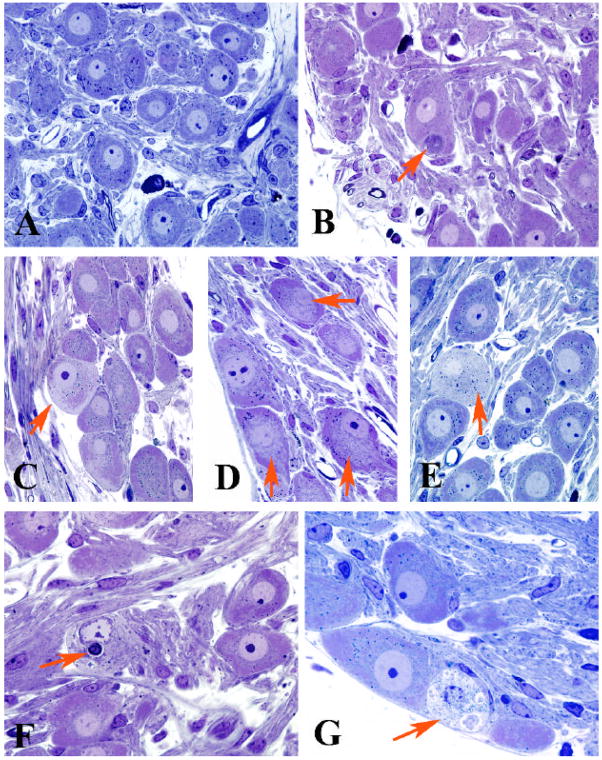
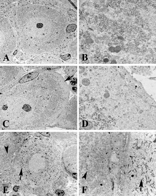
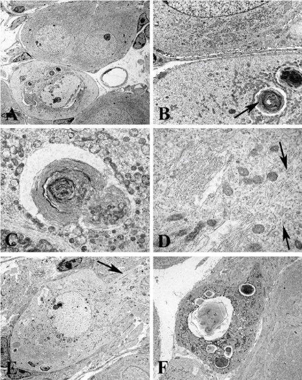
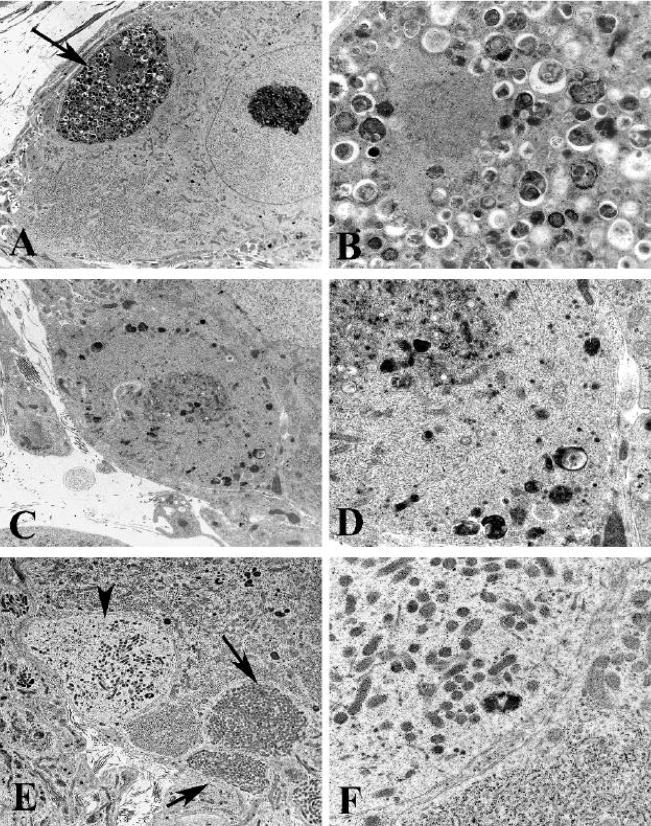
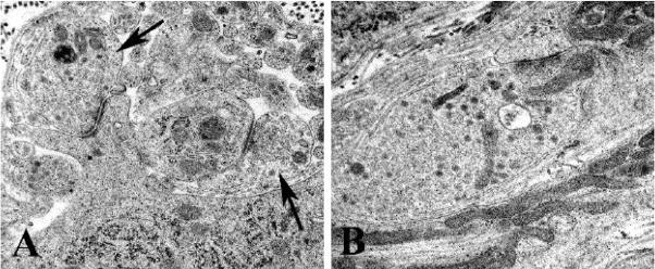
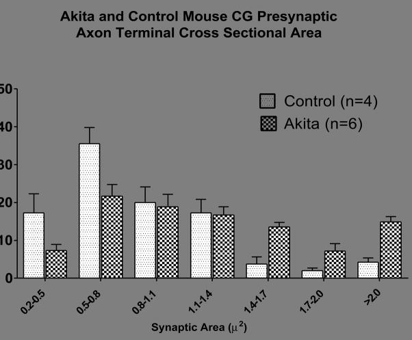
Diabetic autonomic neuropathy is a debilitating, poorly studied complication of diabetes. Our previous studies of non-obese diabetic (NOD) and related mouse models identified rapidly developing, dramatic pathology in prevertebral sympathetic ganglia; however, once diabetic, the mice did not survive for extended periods needed to examine the ability of therapeutic agents to correct established neuropathy. In the current manuscript we show that the Akita (Ins2(Akita)) mouse is a robust model of diabetic sympathetic autonomic neuropathy with unambiguous, spontaneous, rapidly-developing neuropathology which corresponds closely to the characteristic pathology of other rodent models and man. Akita mice diabetic for 2, 4 or 8 months of diabetes progressively developed markedly swollen axons and dendrites ("neuritic dystrophy") in the prevertebral superior mesenteric (SMG) and celiac ganglia (CG). Comparable changes failed to develop in the superior cervical ganglia (SCG) of the Akita mouse or in any ganglia of non-diabetic mice. Morphometric studies demonstrate an overall increase in presynaptic axon terminal cross sectional area, including those without any ultrastructural features of dystrophy. Neurons in Akita mouse prevertebral sympathetic ganglia show an unusual perikaryal alteration characterized by the accumulation of membranous aggregates and minute mitochondria and loss of rough endoplasmic reticulum. These changes result in the loss of a third of neurons in the CG over the course of 8 months of diabetes. The extended survival of diabetic mice and robust pathologic findings provide a clinically relevant paradigm that will facilitate the analysis of novel therapeutic agents on the reversal of autonomic neuropathy.


Figures

Figure 1
Figure 1
Light microscopic appearance of the SMG and CG of control and diabetic Akita mice A) Principal sympathetic neurons surrounded by neuropil with occasional mast cells (control SMG, 5 months of age) B) A typical dystrophic neurite (arrow) is intimately associated with an adjacent cell body (2 month diabetic CG) C–E) Neurons show patchy loss of Nissl substance involving the subplasmalemma (arrow, C), central perinuclear area (arrows, D) and entire perikaryon (arrow, E) [C, 4 month diabetic CG; D, 8 month diabetic SMG; E, 8 month diabetic CG] F,G) Degenerating neurons may contain large membranous aggregates (arrow, F) or disintegrating cytoplasm (arrow, G) [F, 2 month diabetic CG; G, 4 month diabetic SMG] (original magnification A–G, 500X)
Figure 2
Figure 2
Ultrastructural appearance of rough endoplasmic reticulum in the CG of control and Akita mice diabetic for 2 months A,B) Control CG neuron shows a round nucleus with little heterochromatin and a prominent nucleolus with discrete collections of rough endoplasmic reticulum (B). [A, CG, 9 months of age; B, 5 month age SMG] (original magnification: A, 3000X; B, 25000X) C,D) Subplasmalemmal loss of rough endoplasmic reticulum (arrows, C,D) with little additional pathology is a frequent pattern in diabetic Akita neurons. [C,D, 8 month diabetic CG] (original magnification: C, 4000X; D, 30000X) E,F) Complete loss of perikaryal rough endoplasmic reticulum is shown (arrow, E) in comparison to an adjacent neuron with a normal pattern (arrowhead, E). The pale neuronal cytoplasm (arrow, F) contains mitochondria smaller than those of the adjacent normal neuron (arrowhead, F). [4 month diabetic SMG] (original magnification: E, 3000X; F, 12000X)
Figure 3
Figure 3
Ultrastructural appearance of membranous pathology involving the CG of Akita mice diabetic for 2 months A–C) A degenerating neuron containing large membranous aggregates (A), is shown at higher magnification in B and C, and is accompanied by large numbers of minute mitochondria. (original magnification: A, 2500X; B, 12000X; C, 20000X) D) Pale cytoplasm shows large numbers of membranous elements (arrows) without attached ribosomes (original magnification: 40000X) E) Neuronal pallor extends into adjacent proximal dendrite (arrows) (original magnification: 3000X) F) Terminal appearance of degenerating neuron (original magnification: 4000X)
Figure 4
Figure 4
Ultrastructural appearance of neuritic dystrophy in the SMG and CG of Akita mice A,B) Axonal nerve terminal (arrow, A) containing numerous normal and degenerating organelles, and autophagosomes shown at higher magnification in B. [8 month diabetic SMG] (original magnification: A, 5000X; B, 25000X) C,D) Swollen terminal axon with aggregated normal, degenerating and autophagic organelles embedded in disorganized cytoskeleton, seen at higher magnification in D. [4 month diabetic SMG] (original magnification: C, 10000X; D, 25000X) E,F) Dilated dendrites containing large numbers of small mitochondria (arrows, E) and a pale dendritic process (arrowhead, E) containing small mitochondria, tubulovesicular elements and little rough endoplasmic reticulum (F) [8 month diabetic CG] (original magnification: E, 6000X; F, 25000X)
Figure 5
Figure 5
Ultrastructural appearance of typical nerve terminals in control and Akita mouse CG A,B) Typical axonal terminals (arrows, A) forming synapses on a dendrite and a perikaryal spine in a control mouse. The presynaptic nerve terminals of Akita mice (B) are typically larger than those of controls although the material accumulated consists of typical synaptic vesicles and mitochondria [A, 3 months of age; B, 4 month diabetic] (original magnification: A,B, 40000X)
Figure 6
Figure 6
Size-frequency plot of the distribution of presynaptic axon terminal cross sectional areas in the CG of control and Akita mice diabetic for 8 months. Values represent the means + SEM of 4 control and 6 Akita mice ganglia.

References

    1. Appenzeller O, Richardson EP., Jr The sympathetic chain in patients with diabetic and alcoholic polyneuropathy. Neurology. 1966;16:1205–1209. - PubMed
    1. Baloh RH. Mitochondrial dynamics and peripheral neuropathy. Neuroscientist. 2008;14:12–18. - PubMed
    1. Baloh RH, Schmidt RE, Pestronk A, Milbrandt J. Altered axonal mitochondrial transport in the pathogenesis of Charcot-Marie-Tooth disease from mitofusin 2 mutations. J Neurosci. 2007;27:422–430. - PMC - PubMed
    1. Barber AJ, Antonetti DA, Kern TS, Reiter CE, Soans RS, Krady JK, Levison SW, Gardner TW, Bronson SK. The Ins2Akita mouse as a model of early retinal complications in diabetes. Invest Ophthalmol Vis Sci. 2005;46:2210–2218. - PubMed
    1. Budzilovich GN. Diabetic neuropathy complex. Virchows Arch A Pathol Anat. 1970;350:105–122. - PubMed

Publication types

MeSH terms

LinkOut - more resources