Review
. 2009 Apr;1793(4):726-36.
doi: 10.1016/j.bbamcr.2008.11.014. Epub 2008 Dec 9.

Secondary lipid accumulation in lysosomal disease

Affiliations
Review

Secondary lipid accumulation in lysosomal disease

Steven U Walkley et al. Biochim Biophys Acta. 2009 Apr.

Abstract

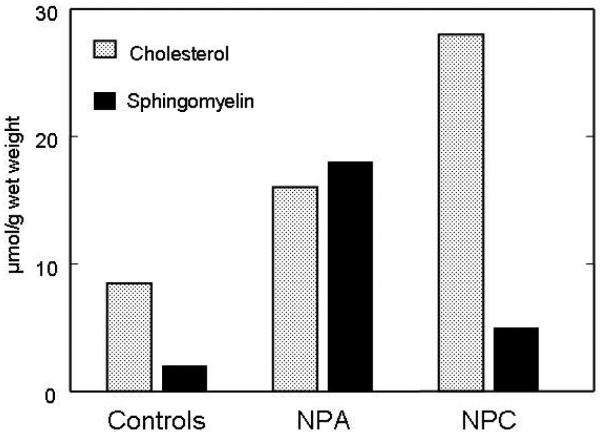
Lysosomal diseases are inherited metabolic disorders caused by defects in a wide spectrum of lysosomal and a few non-lysosomal proteins. In most cases a single type of primary storage material is identified, which has been used to name and classify the disorders: hence the terms sphingolipidoses, gangliosidoses, mucopolysaccharidoses, glycoproteinoses, and so forth. In addition to this primary storage, however, a host of secondary storage products can also be identified, more often than not having no direct link to the primary protein defect. Lipids - glycosphingolipids and phospholipids, as well as cholesterol - are the most ubiquitous and best studied of these secondary storage materials. While in the past typically considered nonspecific and nonconsequential features of these diseases, newer studies suggest direct links between secondary storage and disease pathogenesis and support the view that understanding all aspects of this sequestration process will provide important insights into the cell biology and treatment of lysosomal disease.


Figures

Figure 1
Figure 1
Gangliosides GM3 and GM2 in brain from mouse models of several lysosomal storage diseases. All mice were studied in the laboratory of one of us (MTV) using the same procedure. Results are expressed in nmol ganglioside/g wet weight of tissue. WT: wild type; PSAP: prosaposin deficiency; ctsd: cathepsin D deficiency (CLN10); Hex-0: Hexosaminidase A + B deficiency (GM2-gangliosidosis, Sandhoff variant); NPC1: NPC1 deficiency (Niemann-Pick C); MPSI: α-iduronidase deficiency; MPSIIIb: α-N-acetylglucosaminidase (NAGLU) deficiency; NPA: acid sphingomyelinase deficiency (Niemann-Pick A); Hex-A: hexosaminidase A deficiency (GM2-gangliosidosis, Tay-Sachs variant). Age at study varied from 1 month to 10 months, as indicated; Note that the GM2 level in the Sandhoff mouse at 1 month of age is already higher than in the Tay-Sachs model at 10 months of age.
Figure 2
Figure 2
Gangliosides GM3 and GM2 and cholesterol in two adjacent cerebrocortical neurons in murine MPS IIIA disease. Confocal analysis reveals the subcellular localization of GM2, GM3, and cholesterol. A: GM2 immunofluorescence. B: GM3 immunofluorescence. C: Filipin histochemistry (or unesterified cholesterol). D: Merged image. As is evident, most labeled vesicles in A and B contained either GM2 or GM3 gangliosides, but very few showed significant co-labeling (D). While most cholesterol also appeared to occur independently of ganglioside-containing vesicles, GM3 ganglioside and cholesterol exhibited similar subcellular distribution, as shown by the neuron on left (arrow). Note that the cell on the right in C (and D) does not exhibit significant cholesterol accumulation (it is largely filipin-negative). Scale bar in D equals 10 microns and applies to A–D. (From ref. 34, used with permission).
Figure 3
Figure 3
Filipin histochemical staining for unesterified cholesterol in cerebrocortical neurons in multiple lysosomal diseases. A: Wt. (12 weeks old) B: MPS IIIA disease (12 weeks old). C: Niemann-Pick disease type C (8 weeks old). D: GM1 gangliosidosis (12 weeks old). Note that neurons in Wt brain exhibit no significant filipin labeling of somata, whereas in each of the lysosomal diseases there is substantial filipin labeling of individual neurons. In MPS IIIA disease, some neurons are clearly more affected than others, whereas in Niemann-Pick C and GM1 gangliosidosis all neurons are positive. Filipin staining in GM1 was substantial and appeared to exceed that of Niemann-Pick C in late stage disease. Calibration bar in C equals 12 µm and applies to all.
Figure 4
Figure 4
Lipid storage in liver from 20 week-old foetuses with Niemann-Pick type A (NPA) (3 cases) and type C (NPC) (3 cases) diseases. This was compared to concentrations in foetuses with lysosomal disorders not affecting the liver (controls, 5 cases). All results are expressed as mean values for each group. In NPA, the primary stored lipid, sphingomyelin, is already very elevated, and is accompanied by a prominent secondary increase of cholesterol. In NPC, massive cholesterol storage occurs, with a modest sphingomyelin increase.

References

    1. Platt FM, Walkley SU. Lysosomal Defects and Storage. In: Platt FM, Walkley SU, editors. Lysosomal Disorders of the Brain. Oxford University Press; Oxford: 2004. pp. 32–49.
    1. Cumings JN. Abnormalities in lipid metabolism in two members of a amily with Niemann-Pick disease. In: Aronson SM, Volk BW, editors. Cerebral lipidoses. Academic Press; New York: 1962. pp. 171–178.
    1. Kamoshita S, Aron AM, Suzuki K, Suzuki K. Infantile Niemann-Pick disease. A chemical study with isolation and characterization of membranous cytoplasmic bodies and myelin. Am. J. Dis. Child. 1969;117:379–394. - PubMed
    1. Martin JJ, Philippart M, Van Hauwaert J, Callahan JW, Deberdt R. Niemann-Pick disease (Crocker's group A). Late onset and pigmentary degeneration resembling Hallervorden-Spatz syndrome. Arch. Neurol. 1972;27:45–51. - PubMed
    1. Brunngraber EG, Berra B, Zambotti V. Altered levels of tissue glycoproteins, gangliosides, glycosaminoglycans and lipids in Niemann-Pick's disease. Clin. Chim. Acta. 1973;48:173–181. - PubMed

Publication types