. 2008 Dec 26;32(6):849-61.
doi: 10.1016/j.molcel.2008.11.018.

Structure of a complete integrin ectodomain in a physiologic resting state and activation and deactivation by applied forces

Affiliations

Structure of a complete integrin ectodomain in a physiologic resting state and activation and deactivation by applied forces

Jianghai Zhu et al. Mol Cell. .

Abstract

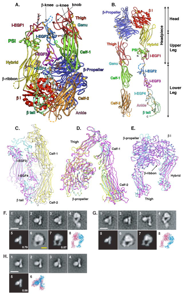
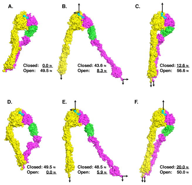
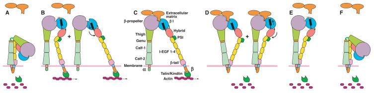
The complete ectodomain of integrin alpha(IIb)beta(3) reveals a bent, closed, low-affinity conformation, the beta knee, and a mechanism for linking cytoskeleton attachment to high affinity for ligand. Ca and Mg ions in the recognition site, including the synergistic metal ion binding site (SyMBS), are loaded prior to ligand binding. Electrophilicity of the ligand-binding Mg ion is increased in the open conformation. The beta(3) knee passes between the beta(3)-PSI and alpha(IIb)-knob to bury the lower beta leg in a cleft, from which it is released for extension. Different integrin molecules in crystals and EM reveal breathing that appears on pathway to extension. Tensile force applied to the extended ligand-receptor complex stabilizes the closed, low-affinity conformation. By contrast, an additional lateral force applied to the beta subunit to mimic attachment to moving actin filaments stabilizes the open, high-affinity conformation. This mechanism propagates allostery over long distances and couples cytoskeleton attachment of integrins to their high-affinity state.


Figures

Figure 1
Figure 1
The αIIb β3 crystal structure. A. Cartoon diagram of molecule 1 in αIIb β3 crystals. Ca and Mg ions are shown as gold and silver spheres, respectively. Disulfides are shown as gold sticks and glycans are displayed as thinner sticks with grey carbons. C and N-termini are shown as small spheres. Loops with missing density are shown as dashes. B. A model of αIIb β3 extended by torsion at the α and β-knees. C-E. Superpositions of molecules 1 and 2 of αIIb β3 and αV β3 (Xiong et al., 2004) showing breathing. C. A view showing variation in the distance of the lower α-leg from the lower β-leg, opening its cleft, and variation in the lower β-leg: αIIb β3 molecule 1 (αIIb grey, β3 cyan) and αV β3V yellow, β3 magenta). D. A view of the α-subunit only, rotated about 90° from the view in C, showing variation in the distance of the lower α-leg from the upper α-headpiece: αIIb β3 molecule 1 (yellow) and molecule 2 (cyan); αV β3 (magenta). E. The headpieces of αIIb β3 molecule 1 (cyan) and αVβ3 (magenta), showing breathing at the β I/hybrid domain interface. All figures are made with PYMOL. F-H. Negatively stained αIIb β3 EM projection averages. F. αIIb β3 with a C-terminal coiled-coil clasp. G. αIIb β3 with the clasp removed. H. αIIb β3 disulfide-bonded near the C-termini of the β-tail and calf-2 domains. Panels 1–4 show representative class averages. Panel 5 shows the 20 Å resolution-filtered αIIb β3 crystal structure projection that best cross-correlates with panel 1. Panel 6 in F and G shows the masked headpiece region from panel 4, and panel 7 shows the corresponding best-correlated αIIb β3 headpiece crystal structure projection. Ribbon diagrams in panels 6 and 8 are in the same orientation (although enlarged) as the projections to their left. Numbers in panels 5 and 7 are normalized cross-correlation coefficients. White and yellow scale bars are 100 and 50 Å, respectively.
Figure 2
Figure 2
Integrin leg domains. A. The knee and lower β-leg of αIIb β3. Dashes mark gimbal flexion positions. B. Superposition using SSM (Krissinel and Henrick, 2004) of αIIb β3 I-EGF domains 1–4 in the same color scheme as in A. C. Superposition using I-EGF2 of I-EGF 1 and 2 module pairs. Domains from αIIb β3 are in red, and those from β2 fragments (Shi et al., 2007) are in cyan and grey. D-F. Superposition of αIIb (yellow) and αV (magenta) (Xiong et al., 2004) thigh (D), calf-1 (E), and calf-2 (F) domains.
Figure 3
Figure 3
Metal ion rearrangements in β I domain activation. A. Superposition of headpieces from our unliganded-closed structure and liganded-open αIIb β3 (Springer et al., 2008). The β I and hybrid domains are yellow (open) and magenta (closed) while PSI and I-EGF1 domains are red and green, respectively. The α-headpieces are cyan (open) and grey (closed). B. Enlarged view of β I domains with major differences in yellow (open) and magenta (closed). C and D. β I domain metal coordination sites in unliganded-closed αIIb β3 (C) and liganded-open αIIb β3 (D). Ca (gold) and Mg (green) ions are large spheres; waters (red or pink) are smaller spheres. N atoms are blue and O atoms are red or pink. Metal coordination and hydrogen bonds are dashed. The loop bearing M335 moves far away in (D). E. Superposition at the β I MIDAS. F. Superposition at the α I MIDAS of unliganded-closed (PDB code 1LFA) and liganded open αL (PDB code 1T0P), in the same orientation as the β I MIDAS in D. In C-F, carbons for unliganded-closed and liganded-open integrins and for ligands are wheat, grey, and cyan, respectively. G and H. Electrostatic potential surfaces at the unliganded (G) and liganded (H) binding sites.
Figure 4
Figure 4
Regulation of integrin conformation by tensile force. Molecular surfaces show αIIb in yellow and β3 in magenta with hybrid domain in green. Fibrinogen peptide ligand is shown in cyan as Cα spheres. A and D are starting models. B and C are derived from A, and E and F from D, after applying tensile forces (arrows) in steered molecular dynamics simulations to αIIb and β3 C-terminal and ligand N-terminal atoms shown as large spheres. Models in A-F are aligned by superposition on the β-propeller and β I domains. Numbers show the distance after superposition of C-terminal residue 433 of the hybrid domain from the closed (A) and open (D) conformations. The underlined distance shows the conformation that models most closely resemble.
Figure 5
Figure 5
The integrin cycle. A. In the bent conformation, integrins have low affinity for ligand. B. At sites where actin filaments are formed, the integrin β subunit cytoplasmic domain binds through talin or kindlins. Lateral translocation on the cell surface and buffetting cause integrin extension. Both open and closed headpiece conformations are putatively present. C. Binding to an immoblized extracellular ligand greatly increases the lateral force, and markedly favors the high-affinity, open headpiece conformation. D. Disassembly of the actin cytoskeleton removes the lateral force. Tensile force between the ligand and the integrin cytoplasmic domains favors the closed headpiece conformation, and ligand dissociation. E. Ligand dissociates, further favoring the closed headpiece conformation. F. In the absence of ligand and tensile force, the bent conformation is favored, completing the cycle, and the integrin returns to the same state as shown in (A).

References

    1. Adair BD, Yeager M. Three-dimensional model of the human platelet integrin αIIb β3 based on electron cryomicroscopy and x-ray crystallography. Proc Natl Acad Sci U S A. 2002;99:14059–14064. - PMC - PubMed
    1. Alon R, Dustin ML. Force as a Facilitator of Integrin Conformational Changes during Leukocyte Arrest on Blood Vessels and Antigen-Presenting Cells. Immunity. 2007;26:17–27. - PubMed
    1. Arnaout MA, Mahalingam B, Xiong JP. Integrin structure, allostery, and bidirectional signaling. Annu Rev Cell Dev Biol. 2005;21:381–410. - PubMed
    1. Astrof NS, Salas A, Shimaoka M, Chen JF, Springer TA. Importance of force linkage in mechanochemistry of adhesion receptors. Biochemistry. 2006;45:15020–15028. - PMC - PubMed
    1. Beglova N, Blacklow SC, Takagi J, Springer TA. Cysteine-rich module structure reveals a fulcrum for integrin rearrangement upon activation. Nat Struct Biol. 2002;9:282–287. - PubMed

Publication types

LinkOut - more resources