. 2009 Jan 6;15(1):35-44.
doi: 10.1016/j.ccr.2008.11.012.

Hypoxia-induced lysyl oxidase is a critical mediator of bone marrow cell recruitment to form the premetastatic niche

Affiliations

Hypoxia-induced lysyl oxidase is a critical mediator of bone marrow cell recruitment to form the premetastatic niche

Janine T Erler et al. Cancer Cell. .

Abstract

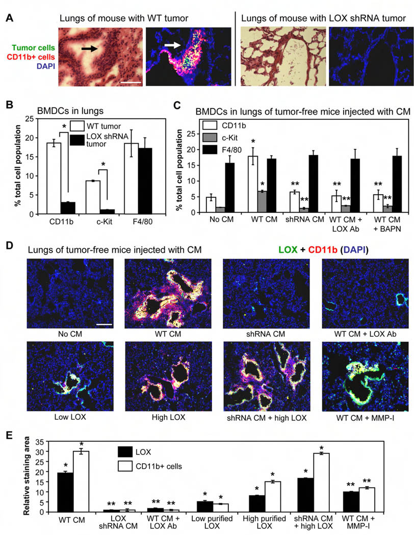
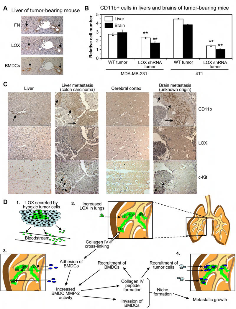
Tumor cell metastasis is facilitated by "premetastatic niches" formed in destination organs by invading bone marrow-derived cells (BMDCs). Lysyl oxidase (LOX) is critical for premetastatic niche formation. LOX secreted by hypoxic breast tumor cells accumulates at premetastatic sites, crosslinks collagen IV in the basement membrane, and is essential for CD11b+ myeloid cell recruitment. CD11b+ cells adhere to crosslinked collagen IV and produce matrix metalloproteinase-2, which cleaves collagen, enhancing the invasion and recruitment of BMDCs and metastasizing tumor cells. LOX inhibition prevents CD11b+ cell recruitment and metastatic growth. CD11b+ cells and LOX also colocalize in biopsies of human metastases. Our findings demonstrate a critical role for LOX in premetastatic niche formation and support targeting LOX for the treatment and prevention of metastatic disease.


Figures

Figure 1
Figure 1. LOX secreted from hypoxic tumor cells co-localizes with CD11b+ cells in the lungs and increases CD11b+ cell recruitment and invasion
(A) Nude mice were orthotopically implanted with 107 wild-type (Wt) or LOX shRNA-expressing MDA-MB-231 human breast tumor cells. Lungs were excised 6 weeks later, and frozen serial sections were stained with either H&E or with pan-cytokeratin (green) to identify tumor cells and CD11b (red) to identify myeloid cells. Arrows indicate pulmonary cell clusters (foci). Scale bar = 75µm. (B) Lungs from mice bearing Wt or LOX shRNA-expressing tumors were homogenized and analyzed by flow cytometry for numbers of CD11b+ myeloid cells, c-Kit+ (CD117) myeloid progenitor cells, or F4/80+ mature macrophages. Data are mean ± SEM. * = p<0.05. (C) Tumor-free mice were injected with the indicated CM daily for 3 weeks. Homogenized lungs were analyzed for CD11b, c-Kit, and F4/80 positive cells by flow cytometry. LOX Ab = LOX-specific antibody; BAPN = small molecule inhibitor of LOX. Data are mean ± SEM. * = p<0.05 relative to control; ** = p<0.05 relative to mice injected with Wt CM. (D) Merged immunofluorescent staining of LOX (green) and CD11b+ cells (red) in representative lung sections from tumor-free mice injected daily for 3 weeks with the indicated CM. Co-localization is indicated in yellow. Low/high LOX = relative concentration of purified LOX; MMP-I = matrix metalloproteinase inhibitor. Scale bar = 150µm. (E) Image analysis of lung sections from mice in Figure 1D. Data indicate the relative area of LOX (green) or CD11b (red) staining relative to control mice. Data are mean ± SEM. * = p<0.05 relative to control; ** = p<0.05 relative to mice injected with Wt CM.
Figure 2
Figure 2. LOX secreted from hypoxic tumor cells promotes BMDC invasion by cross-linking collagen IV, increasing BMDC adhesion, and enhancing MMP-2 activity of the invading BMDCs
(A) Matrigel-coated wells were incubated with the indicated additives for 24hr; Matrigel cross-linked with glucose was included for comparison. Solutions were removed and CD11b+ cells isolated from murine whole bone marrow were added. Numbers of CD11b+ cells remaining in solution were quantified after 2.5hr. Data are mean ± SEM. * = p<0.05 relative to control; ** = p<0.05 relative to matrix pre-incubated with LOX. (B) Naïve mouse lung tissue was excised and a 2cm3 piece was incubated in serum-free media containing either LOX or glucose for 6hr. Media was changed, CD11b+ cells were added, and the numbers of cells remaining in solution after 12hr were quantified. Data are mean ± SEM. (C) Lung tissue from B was frozen, sectioned, and stained for LOX (green) and CD11b+ cells (red). Scale bar = 300µm. (D) Gelatin zymography showing MMP-2 activity of monocytes in contact with Matrigel or collagen IV pre-incubated with the indicated CM. (E) Gelatin zymography showing MMP-2 activity of freshly harvested BMDCs in contact with collagen IV pre-incubated with the indicated CM. (F) Matrigel filters were incubated with the indicated CM or purified protein for 24hr. The CM was then removed and freshly harvested whole murine bone marrow cells were allowed to invade through the “modified” Matrigel. BMDCs that invaded through the modified Matrigel were also stained for CD11b and c-Kit. Data are mean ± SEM. * = p<0.05 relative to control; ** = p<0.05 relative to matrices pre-incubated with Wt CM. (G) Matrigel filters were pre-incubated as in 2F, and invasion of isolated CD11b+ cells or c-Kit+ cells through the modified matrix was quantified. Data are mean ± SEM. * = p<0.05 relative to control; ** = p<0.05 relative to matrices pre-incubated with LOX. (H) Mice with wild-type bone marrow or MMP-2 knockout bone marrow were injected daily with the indicated CM for 3 weeks prior to flow cytometry analysis of lungs for CD11b, c-Kit, or F4/80 positive cells. Data are mean ± SEM. * = p<0.05 relative to no CM mice; ** = p<0.05 relative to mice with Wt bone marrow injected with Wt CM. (I) Immunofluorescent staining for LOX, CD11b+ cells, and MMP-2 in representative frozen serial sections of lungs from mice with wild-type or MMP-2 knockout bone marrow injected with Wt CM. Scale bar = 75µm.
Figure 3
Figure 3. Collagen IV remodeling mediated by LOX and BMDC MMP-2 activity promotes pulmonary metastatic growth
(A) ELISA to detect collagen IV remodeling via peptide formation. Collagen IV peptides exogenously added to media (white bars) are provided for comparison. Matrigel pre-incubated for 24hr with the indicated additives (grey bars) were subsequently contacted with CD11b+ cells for 24hr. Peptides released into the surrounding media were quantified with the ELISA. Plasma samples from the indicated tumor-bearing mice (black bars) were also analyzed. Data are mean ± SEM. * = p<0.05 relative to control; ** = p<0.05 relative to matrices pre-incubated with LOX (grey) or relative to Wt tumor-bearing mice (black). (B) Sections of lungs from Wt tumor-bearing mouse illustrating loss of collagen IV antibody epitope in some areas through LOX-mediated collagen IV remodeling. Collagen I staining was not affected. Scale bar = 150µm. (C) Numbers of isolated CD11b+ cells or c-Kit+ cells invading through naïve Matrigel toward collagen IV peptides in the bottom of the transwell. Data are mean ± SEM. * = p<0.05 relative to control (no peptides). (D) Flow cytometric quantification of CD11b+ cells and tumor cells (human pan-cytokeratin-positive) in lungs of mice bearing LOX shRNA-expressing tumors. Mice were “pre-conditioned” by injection of Wt CM or purified LOX protein for 2 weeks after tumor implant. Lungs were harvested 6 weeks after tumor implant. Data are mean ± SEM. * = p<0.05 relative to control mice; ** = p<0.05 relative to mice pre-conditioned with LOX protein. (E) Flow cytometric quantification of CD11b+ cells and tumor cells in lungs of mice with LOX shRNA-expressing tumors treated with the indicated solutions. Data are mean ± SEM. Clod = clodronate; * = p<0.05 relative to control shRNA tumor-bearing mice; ** = p<0.05 relative to shRNA tumor-bearing mice injected with LOX protein. (F) Same experiment as E, using 4T1 murine mammary tumor cells in Balb/c mice. Pulmonary cell foci (clusters) were quantified from H&E-stained lung tissue. Data are mean ± SEM. * = p<0.05 relative to control 4T1 shRNA tumor-bearing mice; ** = p<0.05 relative to 4T1 shRNA tumor-bearing mice injected with LOX protein. (G) Flow cytometry analysis for CD11b+ cells in lungs of tumor-free Balb/c mice or Balb/c mice bearing LOX shRNA-expressing 4T1 tumors injected with the indicated CM daily for 3 weeks. Pulmonary cell foci were quantified from H&E-stained lung tissue. Data are mean ± SEM. * = p<0.05 relative to control mice; ** = p<0.05 relative to mice injected with Wt CM. (H) Lungs of Balb/c mice implanted with LOX shRNA-expressing 4T1 tumors and injected with the indicated CM daily for 3 weeks. Arrows indicate macroscopic lung metastases.
Figure 4
Figure 4. CD11b+ cells and LOX are associated with liver and brain metastases in patients
(A) Serial sections from liver tissue of Wt tumor-bearing mouse stained for fibronectin, LOX, and BMDCs. Scale bar = 50µm. (B) Flow cytometry analysis for CD11b+ cells in livers and brains of Nude mice bearing MDA-MB-231 human breast tumors or Balb/c mice with 4T1 murine mammary tumors. Data are mean ± SEM. ** = p<0.05 relative to Wt tumor-bearing mice. (C) Tissue microarrays (TMAs) of clinical metastases stained for CD11b+ cells, LOX, or c-Kit+ cells. Samples from normal cerebral cortex and liver are provided as negative controls. Metastatic and normal TMAs were stained simultaneously, and were photographed with identical microscope and camera settings. Arrows indicate regions of CD11b+ cells or c-Kit+ cells. Scale bar = 150µm. (D) (1) Hypoxic primary tumor cells secrete LOX into the bloodstream. (2) LOX accumulates in the lungs of tumor-bearing mice and cross-links collagen IV. (3) Adhesion of CD11b+ cells to cross-linked matrix increases BMDC MMP-2 activity. Collagen IV remodeling by LOX and MMP-2 leads to peptide formation, invasion of CD11b+ cells, and increased recruitment of BMDCs. (4) LOX-dependent formation of the pre-metastatic niche enhances metastatic growth.

References

    1. Ahn GO, Brown JM. Matrix metalloproteinase-9 is required for tumor vasculogenesis but not for angiogenesis: role of bone marrow-derived myelomonocytic cells. Cancer Cell. 2008;13:193–205. - PMC - PubMed
    1. Cairns RA, Khokha R, Hill RP. Molecular mechanisms of tumor invasion and metastasis: an integrated view. Curr Mol Med. 2003;3:659–671. - PubMed
    1. Cameron JD, Skubitz AP, Furcht LT. Type IV collagen and corneal epithelial adhesion and migration. Effects of type IV collagen fragments and synthetic peptides on rabbit corneal epithelial cell adhesion and migration in vitro. Invest Ophthalmol Vis Sci. 1991;32:2766–2773. - PubMed
    1. Caniggia I, Mostachfi H, Winter J, Gassmann M, Lye SJ, Kuliszewski M, Post M. Hypoxia-inducible factor-1 mediates the biological effects of oxygen on human trophoblast differentiation through TGFbeta(3) J Clin Invest. 2000;105:577–587. - PMC - PubMed
    1. Condeelis J, Pollard JW. Macrophages: obligate partners for tumor cell migration, invasion, and metastasis. Cell. 2006;124:263–266. - PubMed

Publication types

MeSH terms