. 2009 Jan 6;15(1):45-56.
doi: 10.1016/j.ccr.2008.12.006.

Malignant astrocytomas originate from neural stem/progenitor cells in a somatic tumor suppressor mouse model

Affiliations

Malignant astrocytomas originate from neural stem/progenitor cells in a somatic tumor suppressor mouse model

Sheila Alcantara Llaguno et al. Cancer Cell. .

Erratum in

  • Cancer Cell. 2009 Mar 3;15(3):240

Abstract

Malignant astrocytomas are infiltrative and incurable brain tumors. Despite profound therapeutic implications, the identity of the cell (or cells) of origin has not been rigorously determined. We previously reported mouse models based on conditional inactivation of the human astrocytoma-relevant tumor suppressors p53, Nf1, and Pten, wherein through somatic loss of heterozygosity, mutant mice develop tumors with 100% penetrance. In the present study, we show that tumor suppressor inactivation in neural stem/progenitor cells is both necessary and sufficient to induce astrocytoma formation. We demonstrate in vivo that transformed cells and their progeny undergo infiltration and multilineage differentiation during tumorigenesis. Tumor suppressor heterozygous neural stem/progenitor cultures from presymptomatic mice show aberrant growth advantage and altered differentiation, thus identifying a pretumorigenic cell population.


Figures
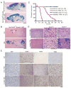
Figure 1
Figure 1. Tumor Suppressor Inactivation in Nestin-expressing Neural Stem/Progenitor Cells Induces High Grade Astrocytoma Formation
A–B. Nestin-creERT2 transgenic mice possess cre recombinase activity in the neural stem/progenitor niches. A. Representative images of X-gal-stained adult brain sections show that cre activity of nestin-creERT2;R26-lacZ mice treated with tamoxifen at E13.5 recapitulates that of the hGFAP-cre transgenic mice. Scale bar, 2 mm. B. Representative images of X-gal-stained brain sections at 6 months post-injection (MPI) reveal that tamoxifen induction at 4 weeks of age mainly restricted cre activity to neural stem cells and their progeny in the subventricular zone (SVZ), rostral migratory stream (RMS) and olfactory bulb (OB), as well as in the subgranular zone and granule cell layer of the dentate gyrus in the hippocampus (HP). Transgene expression is also found in the cerebellum. No staining is found in the adult cortex (CTX) or striatum (ST), or in vehicle-treated mouse brains. Scale bar, 2 mm. C. Mice with induced tumor suppressor inactivation (nestin-creERT2;Nf1flox/+;p53flox/flox;Ptenflox/+ or nestin-creERT2;Nf1flox/flox;p53flox/flox) by tamoxifen treatment (TMX) have shortened survival compared to vehicle-treated (Veh) mice. Kaplan-Meier survival curves of nestin-creERT2 mice with tumor suppressor inactivation induced at either E13.5 or 4 weeks of age show median survivals of 16 weeks and 46 weeks, respectively. Red arrow indicates timing of adult injection. D. Histologically identifiable high grade astrocytomas develop in inducible mutant mice. Representative H&E-stained brain sections reveal formation of brain tumors in nestin-creERT2 mice with tumor suppressor inactivation induced at either E13.5 or 4 weeks of age. Grade III or IV astrocytomas with characteristic features of nuclear atypia, mitoses (arrowheads), and necrosis (N) are shown. Scale bars, 100 μm. E. Tumors express traditional markers of astrocytomas, including Ki67, Gfap, nestin, and Olig2. Scale bar, 100 μm.
Figure 2
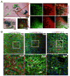
Figure 2. Cancer-initiating Cells Exhibit Infiltration and Differentiation During Tumor Development
A. β-galactosidase-positive mutant cells infiltrate into adjacent brain structures, away from their normal neural stem/progenitor niches. A representative X-gal-stained brain tumor section from nestin-creERT2;Nf1flox/+;p53flox/flox;Ptenflox/+;R26-lacZ mouse 8 months after tumor suppressor inactivation shows tumor cells found outside their original locations in the SVZ/SGZ niches (compared to Figure 1B). β-galactosidase-positive tumor cells, shown here expressing Gfap, nestin, and PDGFRα, are found elsewhere in the forebrain, such as the cortex and striatum. B. Marked astrocytoma cells expressing multi-lineage markers are found in the tumor bulk. β-galactosidase-positive tumor cells in inducible mutant mice, shown here in the thalamus, express markers of neuronal (Tuj1), and glial (S100β and APC) differentiation. Toto-3 marks cell nuclei.
Figure 3
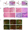
Figure 3. Stereotactic Viral cre-mediated Targeting of the SVZ Induces Astrocytoma Formation Whereas Targeting of Non-neurogenic Regions Does Not
A. Schema of the viral cre injection experiments and representative X-gal staining images. Right, cre adenovirus targeting of the SVZ of R26-lacZ reporter mice marks neural stem/progenitors and its progeny in the RMS (arrowheads) and OB. Left, cre virus injection into non-neurogenic regions such as the striatum causes localized labeling at the site of injection but no labeling at the RMS or OB. Scale bar, 2 mm. B. SVZ-targeted mutant mice develop histologically identifiable high grade astrocytomas. Cre adenovirus was stereotactically injected into the SVZ of tumor suppressor floxed mice (Nf1flox/flox;p53flox/flox, Nf1flox/flox;p53flox/− or Nf1flox/+;p53flox/flox;Ptenflox/+). Analysis of injected mouse brains by H&E staining shows infiltrative tumor cells in SVZ-injected mutant mice (cre→SVZ), whereas mice injected in other non-neurogenic brain regions do not develop tumors (cre→other). Tumors from SVZ-targeted mutant mice show classic histopathologic characteristics of high grade astrocytomas, including tumor (T) invasion into normal (non-tumor, NT) regions, nuclear atypia and mitosis (arrowhead), microvascular proliferation (MP), and necrosis (N). Scale bars, 200 μm. C. Tumors from SVZ-targeted tumor suppressor floxed mice with R26-lacZ reporter show characteristic expression of astrocytoma-associated markers Ki67, Gfap, and nestin. Scale bars, 200 μm.
Figure 4
Figure 4. Tumors arising from SVZ-targeted mutant mice show infiltration and spontaneous differentiation
A. Representative X-gal stained brain sections from mutant mice targeted with cre adenovirus into the SVZ at early postnatal and adult ages show massive tumor formation, extensive infiltration and migration of tumor cells within the brain parenchyma, as well as labeling of the SVZ-RMS-OB axis. LacZ-positive tumor (T) and lacZ-negative non-tumor (NT) regions were dissected out for PCR genotyping, which showed increased recombination of tumor suppressors, in this case, of floxed Nf1 alleles in tumors (G=tail genomic DNA, F=floxed, Δ=recombined, and +=WT=wild type alleles). B. X-gal stained brain sections from mice targeted in non-neurogenic regions such as striatum show localized staining at the site of injection and along the needle tract but no tumor formation. PCR genotyping of lacZ-positive (lacZ+) and lacZ-negative (lacZ−) regions confirms successful recombination of floxed alleles in lacZ+ samples. Scale bar, 2 mm. C. Tumor cells from virus-injected mutant brains exhibit multi-lineage differentiation. Representative immunostaining images of high grade astrocytomas from SVZ-targeted tumor suppressor floxed mice with R26-lacZ reporter injected at early postnatal and adult ages show expression of mature differentiation markers in a subset of β-galactosidase-positive tumor cells, including Gfap for astrocytes, myelin basic protein (MBP) for oligodendrocytes and NeuN for neurons. The majority of marker-positive cells within the tumor bulk, in this case, NeuN-positive cells, were also Pten-negative. A small number of β-galactosidase-positive tumor cells near the cortex were positive for calbindin, a marker of a subset of differentiated OB neurons derived from the SVZ neural stem/progenitor cells. Scale bar, 200 μm. Insets show a β-galactosidase/calbindin double-positive cell. Scale bar, 10 μm.
Figure 5
Figure 5. Pre-tumorigenic SVZ Neural Stem/Progenitor Cells Exhibit Growth Advantage and Altered Differentiation
A. SVZ neurospheres from pre-symptomatic mutant mice exhibit hypertrophy. SVZ cells of 4-week-old mice were cultured as neurospheres. As shown in representative images, Nf1/p53- (Mut3:hGFAP-cre;Nf1flox/+;p53/+) or Nf1/p53/Pten- (Mut4:hGFAP-cre;Nf1flox/+;p53/+;Ptenflox/+) deficient neurospheres were larger than those from wild type (WT). Scale bar, 200 μm. B. Neurosphere proliferation was measured by two hours of bromodeoxyuridine (BrdU) pulsing on day three cultures and subsequent FACS analysis. Mut3 and Mut4 cells had significantly increased S phase, compared to wt cells (p<0.05). C. Neurosphere cell death was measured by Annexin V labeling and subsequent FACS analysis. Mut3 or Mut4 cells undergo significantly reduced cell death compared to wt cells. 7-amino-actinomycin (7-AAD) is a vital dye. D. SVZ neurospheres from pre-symptomatic mutant mice show increased self-renewal potential, as shown by methylcellulose assay compared to wt (**p<0.005). E. Quantification of differentiated cells reveals significant increase in Tuj1- or nestin-positive cells and significant decrease in Gfap-positive cells in Mut3 and Mut4 cultures compared to those of wt control (* p<0.05, **p< 0.005). All error bars are +/− SEM. F. Neurosphere cultures from pre-symptomatic mutant mice exhibit retention of heterozygosity of tumor suppressor genes. Genomic DNA from the ear (E) or neurospheres (N) from four-week-old Mut3 and Mut4 mice was subjected to semi-quantitative PCR genotyping. Representative data for cre, Nf1, p53 and Pten status for neurospheres indicate retention of wild type alleles in all Mut3 and Mut4 neurospheres.

References

    1. Alvarez-Buylla A, Lim DA. For the long run: maintaining germinal niches in the adult brain. Neuron. 2004;41:683–686. - PubMed
    1. Bachoo RM, Maher EA, Ligon KL, Sharpless NE, Chan SS, You MJ, Tang Y, DeFrances J, Stover E, Weissleder R, et al. Epidermal growth factor receptor and Ink4a/Arf: convergent mechanisms governing terminal differentiation and transformation along the neural stem cell to astrocyte axis. Cancer Cell. 2002;1:269–277. - PubMed
    1. Chen J, Kwon C-H, Lin L, Li Y, Parada LF. Inducible Site-Specific Recombination in Neural Stem/Progenitor Cells. Genesis. 2008 in press. - PMC - PubMed
    1. Correa-Cerro LS, Mandell JW. Molecular mechanisms of astrogliosis: new approaches with mouse genetics. J Neuropathol Exp Neurol. 2007;66:169–176. - PubMed
    1. Dai C, Celestino JC, Okada Y, Louis DN, Fuller GN, Holland EC. PDGF autocrine stimulation dedifferentiates cultured astrocytes and induces oligodendrogliomas and oligoastrocytomas from neural progenitors and astrocytes in vivo. Genes Dev. 2001;15:1913–1925. - PMC - PubMed

Publication types

MeSH terms