

Temporoparietal MR imaging measures of atrophy in subjects with mild cognitive impairment that predict subsequent diagnosis of Alzheimer disease
- PMID: 19112067
- PMCID: PMC2656417
- DOI: 10.3174/ajnr.A1397
Temporoparietal MR imaging measures of atrophy in subjects with mild cognitive impairment that predict subsequent diagnosis of Alzheimer disease
Abstract
Background and purpose: Mild cognitive impairment (MCI) represents a transitional state between normal aging and Alzheimer disease (AD). Our goal was to determine if specific temporoparietal regions can predict the time to progress from MCI to AD.
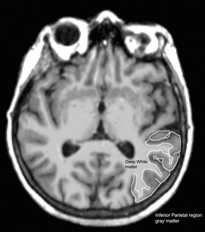
Materials and methods: MR images from 129 individuals with MCI were analyzed to identify the volume of 14 neocortical and 2 non-neocortical brain regions, comprising the temporal and parietal lobes. In addition, 3 neuropsychological test scores were included to determine whether they would provide independent information. After a mean follow-up time of 5 years, 44 of these individuals had progressed to a diagnosis of AD.
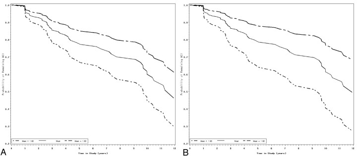
Results: Cox proportional hazards models demonstrated significant effects for 6 MR imaging regions with the greatest differences being the following: the entorhinal cortex (hazard ratio [HR] = 0.54, P < .001), inferior parietal lobule (hazard ratio [HR] = 0.64, P < .005), and middle temporal gyrus (HR = 0.64, P < .004), indicating decreased risk with larger volumes. A multivariable model showed that a combination of the entorhinal cortex (HR = 0.60, P < .001) and the inferior parietal lobule (HR = 0.62, P < .01) was the best predictor of time to progress to AD. A multivariable model reiterated the importance of including both MR imaging and neuropsychological variables in the final model.
Conclusions: These findings reaffirm the importance of the entorhinal cortex and present evidence for the importance of the inferior parietal lobule as a predictor of time to progress from MCI to AD. The inclusion of neuropsychological performance in the final model continues to highlight the importance of using these measures in a complementary fashion.
Figures
References
-
- Petersen R, Smith G, Waring S, et al. Mild cognitive impairment: clinical characterization and outcome. Arch Neurol 1999;56:303–08 - PubMed
-
- Petersen RC, Doody R, Kurz A, et al. Current concepts in mild cognitive impairment. Arch Neurol 2001;58:1985–92 - PubMed
-
- Atiya M, Hyman B, Albert M, et al. Structural magnetic resonance imaging in established and prodromal Alzheimer's disease: a review. Alzheimer Dis Assoc Disord 2003;17:177–95 - PubMed
-
- Chetelat G, Baron JC. Early diagnosis of Alzheimer's disease: contribution of structural neuroimaging. Neuroimage 2003;18:525–41 - PubMed
-
- Kantarci K, Jack C. Neuroimaging in Alzheimer's disease: an evidenced-based review. Neuroimaging Clin N Am 2003;13:197–209 - PubMed
Publication types
MeSH terms
Grants and funding
LinkOut - more resources
Full Text Sources
Medical