. 2009 Apr;85(4):595-605.
doi: 10.1189/jlb.1008631. Epub 2008 Dec 26.

Pivotal Advance: Toll-like receptor regulation of scavenger receptor-A-mediated phagocytosis

Affiliations

Pivotal Advance: Toll-like receptor regulation of scavenger receptor-A-mediated phagocytosis

Eyal Amiel et al. J Leukoc Biol. 2009 Apr.

Abstract

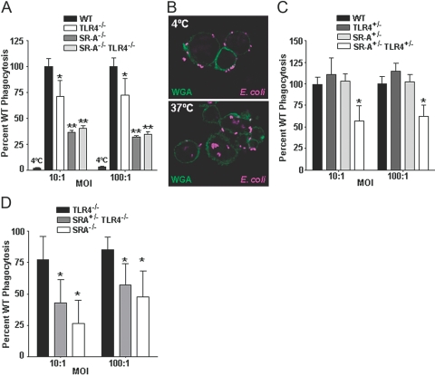
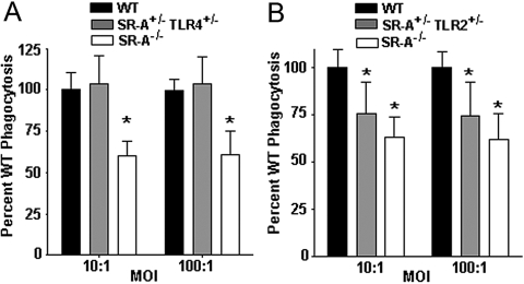
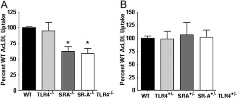
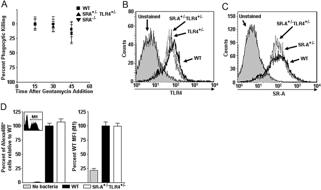
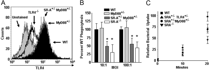
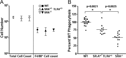
Class-A scavenger receptors (SR-A) and TLR mediate early immune responses against pathogenic bacteria. SR-A and TLR molecules are expressed on phagocytes and interact with common ligands from Gram-negative and Gram-positive bacteria; however, the contribution of TLR activity to SR-A-mediated phagocytosis has not been assessed directly. Herein, we provide genetic and functional evidence that ligand- and TLR-specific stimuli synergize with SR-A to mediate bacterial phagocytosis. Although complete loss of SR-A (SR-A(-/-)) is known to impair bacterial clearance, here we identify the first deficiency attributable to SR-A heterozygosity: SR-A(+/-)TLR4(+/-) cells and mice are impaired significantly in the clearance of Gram-negative Escherichia coli. This phenotype is specific to the TLR signaling event, as SR-A(+/-)TLR4(+/-) cells are not deficient for the clearance of Gram-positive Staphylococcus aureus bacteria, which contain cell-surface TLR2 ligands but lack TLR4 ligands. We demonstrate that this is a global, phagocytic mechanism, regulated independently by multiple TLRs, as analogous to the SR-A(+/-)TLR4(+/-) deficit, SR-A(+/-)TLR2(+/-) cells are impaired for S. aureus uptake. In support of this, we show that SR-A(+/-)MyD88(+/-) cells recapitulate the phagocytosis defect observed in SR-A(+/-)TLR4(+/-) cells. These data identify for the first time that TLR-driven innate immune responses, via a MyD88 signaling mechanism, regulate SR-A-dependent phagocytosis of bacteria. These findings provide novel insights into how innate immune cells control SR-A-mediated trafficking and are the first demonstration that subtle changes in the expression of SR-A and TLRs can substantially affect host bacterial clearance.


Figures

Fig. 1.
Fig. 1.
SR-A+/−TLR4+/− BMDC are impaired in the phagocytosis of E. coli. (A) C57BL/10J (WT), TLR4−/−, SR-A−/−, and SR-A−/−TLR4−/− BMDC were incubated with live E. coli bacteria at 4°C (where noted) or 37°C at the indicated MOIs in a gentamycin protection assay. The mean CFUs of the experimental groups are expressed as a percentage of the mean WT CFU value. TLR4−/− cells were found to be impaired significantly (P<0.05) compared with WT cells. SR-A−/− and SR-A−/−TLR4−/− BMDC were impaired significantly (P<0.01) in the phagocytosis of E. coli compared with WT and TLR4−/− cells but were not significantly different from each other. At 4°C, WT BMDC exhibit >97% reduction in phagocytosis compared with 37°C conditions; n ≥ 12 for all genotypes. (B) C57BL/6 (WT) BMDC were incubated with Alexa647-labeled E. coli for 1 h at 4°C or 37°C. BMDC cell membranes were stained with FITC-labeled wheat germ agglutinin (WGA), and cells were imaged by confocal microscopy. Bacterial internalization was observed at 37°C but not 4°C, consistent with the gentamycin protection assay (A). (C) C57BL/10J, TLR4+/−, SR-A+/−, and SR-A+/−TLR4+/− BMDC were assayed for their quantitative ability to phagocytose live E. coli using a gentamycin protection assay as in (A). TLR4+/− and SR-A+/− BMDC exhibited a similar phagocytic ability as WT cells. However, SR-A+/−TLR4+/− BMDC were impaired significantly (*, P<0.01) in the phagocytosis of E. coli compared with WT BMDC at both MOIs tested. (D) TLR4−/−, SR-A+/−TLR4−/−, and SR-A−/− BMDC were tested for bacterial phagocytosis as in (A and C). BMDC from SR-A+/−TLR4−/− mice exhibited significantly lower levels of phagocytosis than BMDC from TLR4−/− cells; n ≥ 9 for all genotypes. For all graphs, the mean and sd are shown, and statistical significance (P≤0.05) from the WT values (A, C) or the TLR4−/− values (D) is indicated by *, and statistical significance from the WT and TLR4−/− values is indicated by **.
Fig. 2.
Fig. 2.
SR-A+/−TLR2+/−, but not SR-A+/−TLR4+/−, cells are impaired at phagocytosis of the Gram-positive bacteria S. aureus. (A) Gentamycin protection assays were performed on C57BL/10J (WT), SR-A+/−TLR4+/−, and SR-A−/− BMDC, as in Figure 1, with the use of S. aureus bacteria. SR-A+/−TLR4+/− BMDC were comparable with WT cells at the phagocytosis of S. aureus, and SR-A−/− BMDC were impaired significantly (*, P<0.01) compared with WT cells. (B) Gentamycin protection assays were performed on WT, SR-A+/−TLR2+/−, and SR-A−/− BMDC using S. aureus bacteria. SR-A+/−TLR2+/− and SR-A−/− BMDC showed significant impairment in phagocytosis of S. aureus bacteria (*, P<0.001) compared with WT cells. For all graphs, n ≥ 18 for all genotypes, and the means and sd are shown.
Fig. 3.
Fig. 3.
The trafficking deficit exhibited by SR-A+/−TLR4+/− BMDC is specific to SR-A-mediated phagocytosis and does not impair SR-A-mediated endocytic uptake. (A) WT, TLR4−/−, SR-A−/−, and SR-A−/−TLR4−/− BMDC were incubated with Alexa488-labeled AcLDL for 20 min at 37°C and analyzed quantitatively by FACS for endocytic uptake of AcLDL. SR-A−/− and SR-A−/−TLR4−/− BMDC exhibited significant (*, P<0.01) deficits in AcLDL uptake compared with WT cells, and TLR4−/− BMDC were similar to WT BMDC. (B) WT, TLR4+/−, SR-A+/−, and SR-A+/−TLR4+/− BMDC were treated as in A and analyzed for AcLDL uptake by FACS. Single heterozygous genotypes and the double heterozygous BMDC exhibited WT levels of AcLDL uptake.
Fig. 4.
Fig. 4.
SR-A+/−TLR4+/− BMDC exhibit WT levels of bacterial killing and bacterial binding. (A) WT, SR-A+/−TLR4+/−, and SR-A−/− BMDC were incubated with E. coli in a gentamycin protection assay. After gentamycin addition, BMDC from each genotype were harvested every 15 min and lysed to assess the rate of killing of internalized bacteria over the course of the gentamycin protection assay. The graph is shown as percent phagocytic killing over time, and bacterial levels at the first time-point (15 min after gentamycin addition) were set as the baseline by which the later time-points are compared; n ≥ 8 for all genotypes, and mean and sd are shown. No statistical differences were observed among the genotypes. (B) TLR4 surface expression (assayed by FACS) of SR-A+/−TLR4+/− BMDC (dotted line) showed reduced TLR4 expression compared with WT (solid line) cells but identical expression to TLR4+/− cells (dashed line). (C) SR-A surface expression (assayed by FACS) of SR-A+/−TLR4+/− BMDC showed reduced expression of SR-A compared with WT (solid line) cells but identical expression to SR-A+/− cells (dashed line). (D) BMDC were incubated with Alex488-labeled E. coli in the linear range of binding. Gate M1 of the FACS histogram (inset) indicates BMDC that have bound Alexa488 E. coli. Neither the percent of the BMDC population that bound bacteria (number of cells within Gate M1 divided by the total number of cells; left panel) nor the relative number of bacteria bound to the cell surface per BMDC [mean fluorescence intensity (MFI) of Alex488+ BMDC within M1; right panel] was found to differ between WT and SR-A+/−TLR4+/− BMDC. For each parameter, the data are represented as a percentage of mean WT levels.
Fig. 5.
Fig. 5.
SR-A+/−TLR4+/− BMDC exhibit impaired kinetics of bacterial internalization and SR-A+/−MyD88+/− cells recapitulate this defect. (A) FACS histogram showing TLR4 cell-surface expression on BMDC from WT, TLR4−/−, MyD88+/−, and SR-A+/−MyD88+/− mice. MyD88 heterozygosity does not affect cell-surface expression of TLR4. (B) Gentamycin protection assays were performed on WT, MyD88+/−, SR-A+/−MyD88+/−, and SR-A−/− BMDC using E. coli bacteria. SR-A+/−MyD88+/− and SR-A−/− BMDC showed significant impairment in phagocytosis of E. coli (*, P<0.05) compared with WT cells. For all graphs, n ≥ 18 for all genotypes, and the means and sd are shown. (C) Kinetic analysis of bacterial uptake was performed on WT, SR-A+/−TLR4+/−, SR-A+/−MyD88+/−, and SR-A−/− BMDC. Quantitative uptake of bacteria by BMDC was assessed every 10 min using a gentamycin protection assay to determine the relative rates of phagocytosis for the indicated BMDC genotypes. SR-A+/−TLR4+/−, SR-A+/−MyD88+/−, and SR-A−/− BMDC show impairment for the internalization of bacteria as early as 10 min after BMDC-bacteria coincubation, and this deficit is fully apparent by 20 min after bacterial exposure. For all graphs, the means and sd are shown.
Fig. 6.
Fig. 6.
SR-A+/−TLR4+/− mice are impaired at the in vivo phagocytosis of E. coli. WT, SR-A+/−TLR4+/−, and SR-A−/− mice were i.p.-injected with 5 × 10⁶ live E. coli. After 1 h, mice were killed and cells harvested from the peritoneal lavage were treated in a gentamycin protection assay. Relative phagocytosis was determined as a percentage of WT CFU numbers. (A) WT, SR-A+/−TLR4+/−, and SR-A−/− mice showed similar numbers of total cells and F4/80+ cells in peritoneal exudates 1 h after peritoneal injection with live E. coli. Mean and sd are shown. (B) SR-A+/−TLR4+/− mice were impaired significantly (*, P<0.005) for the in vivo uptake of E. coli compared with WT mice, and SR-A−/− mice were impaired even further relative to SR-A+/−TLR4+/− mice (*, P<0.005). Values from individual mice in each group are represented on the graph along with the mean value for each group.

References

    1. Janeway C A, Jr, Medzhitov R. Innate immune recognition. Annu Rev Immunol. 2002;20:197–216. - PubMed
    1. Medzhitov R, Janeway C A., Jr Decoding the patterns of self and nonself by the innate immune system. Science. 2002;296:298–300. - PubMed
    1. Blander J M, Medzhitov R. Regulation of phagosome maturation by signals from Toll-like receptors. Science. 2004;304:1014–1018. - PubMed
    1. Blander J M, Medzhitov R. On regulation of phagosome maturation and antigen presentation. Nat Immunol. 2006;7:1029–1035. - PubMed
    1. Gough P J, Gordon S. The role of scavenger receptors in the innate immune system. Microbes Infect. 2000;2:305–311. - PubMed

Publication types

MeSH terms