. 2009 Mar;296(3):H678-88.
doi: 10.1152/ajpheart.00777.2008. Epub 2008 Dec 26.

Chronic hypoxia enhances 15-lipoxygenase-mediated vasorelaxation in rabbit arteries

Affiliations

Chronic hypoxia enhances 15-lipoxygenase-mediated vasorelaxation in rabbit arteries

Nitin T Aggarwal et al. Am J Physiol Heart Circ Physiol. 2009 Mar.

Retraction in

  • Retraction.
    [No authors listed] [No authors listed] Am J Physiol Heart Circ Physiol. 2014 Aug 15;307(4):H633. doi: 10.1152/ajpheart.zh4-1249-retr.2014. Am J Physiol Heart Circ Physiol. 2014. PMID: 25128501 Free PMC article. No abstract available.

Abstract

15-Lipoxygenase (15-LO-1) metabolizes arachidonic acid (AA) to 11,12,15-trihydroxyeicosatrienoic acids (THETAs) and 15-hydroxy-11,12-epoxyeicosatrienoic acids (HEETA) that dilate rabbit arteries. Increased endothelial 15-LO-1 expression enhances arterial relaxations to agonists. We tested the effect of hypoxia on 15-LO-1 expression, THETA and HEETA synthesis, and relaxations in rabbit arteries. The incubation of rabbit aortic endothelial cells and isolated aortas in 0.7% O(2) increased 15-LO-1 expression. Rabbits were housed in a hypoxic atmosphere of 12% O(2) for 5 days. 15-LO-1 expression increased in the endothelium of the arteries of rabbits in 12% O(2) compared with room air. THETA and HEETA synthesis was also enhanced in aortas and mesenteric arteries. AA hyperpolarized the smooth muscle cells in indomethacin- and phenylephrine-treated mesenteric arteries of hypoxic rabbits from -29.4 +/- 1 to -50.1 +/- 3 mV. The hyperpolarization to AA was less in arteries of normoxic rabbits (from -26.0 +/- 2 to -37 +/- 2 mV). This AA-induced hyperpolarization was inhibited by the 15-LO inhibitor BW-755C. Nitric oxide and prostaglandin-independent maximum relaxations to acetylcholine (79.7 +/- 2%) and AA (38.3 +/- 4%) were enhanced in mesenteric arteries from hypoxic rabbits compared with the normoxic rabbits (49.7 +/- 6% and 19.9 +/- 2%, respectively). These relaxations were inhibited by BW-755C and nordihydroguaiaretic acid. Therefore, hypoxia increased the relaxations to agonists in the rabbit mesenteric arteries by enhancing endothelial 15-LO-1 expression and synthesis of the hyperpolarizing factors THETA and HEETA.


Figures

Fig. 1.
Fig. 1.
15-Lipoxygenase (15-LO-1) protein and mRNA expression in rabbit aortic endothelial cells (RAECs). A: Western immunoblot of 15-LO-1 and β-actin in lysates (40 μg protein). Lysates were prepared from RAECs incubated in normoxic conditions or hypoxic chamber for various times (in hours) and separated on 10% SDS gel. B: 15-LO-1 band density was normalized to the β-actin band density. C: 15-LO-1 mRNA expression in RAECs incubated in the hypoxic chamber quantified by quantitative (q)PCR shown as a change of cycle threshold (ΔCt). Ct values for 15-LO-1 were normalized to Ct values for GAPDH to obtain ΔCt. D: agarose gel separation of the amplified products obtained from qPCR. Values represent means ± SE. *P < 0.05 compared with the value at time 0. RT, reverse transcriptase; T, cDNA template.
Fig. 2.
Fig. 2.
15-LO-1 and platelet endothelial cell adhesion molecule (PECAM) expression in isolated rabbit aortas. Aortic rings incubated in normoxic (N) or hypoxic (H) conditions for various times (in hours). Lysates were prepared and separated on 10% SDS gel. A: Western immunoblot of 15-LO-1 and β-actin in lysates (40 μg protein). B: 15-LO-1 band density was normalized to β-actin band density. C: Western immunoblot of PECAM and β-actin in lysates (40 μg protein). D: PECAM band density was normalized to the β-actin band density. E: 15-LO-1 mRNA expression in aortic rings incubated in normoxic or hypoxic conditions and quantified by qPCR. Ct values for 15-LO-1 were normalized to Ct values for GAPDH to obtain ΔCt. F: agarose gel separation of the amplified products obtained from qPCR. Values represent means ± SE. **P < 0.01 and *P < 0.05 compared with the corresponding values in normoxic arteries.
Fig. 3.
Fig. 3.
15-LO-1 protein and mRNA expression in arteries from normoxic or hypoxic rabbits. A: Western immunoblot of 15-LO-1 and β-actin in lysates (40 μg protein). Lysates were prepared from various arteries from normoxic or hypoxic rabbits and separated on 10% SDS gel. B: 15-LO-1 band density was normalized to the β-actin band density. Values represent means ± SE. ***P < 0.001, **P < 0.01, and *P < 0.05 compared with the corresponding values in normoxic arteries.
Fig. 4.
Fig. 4.
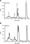
Arachidonic acid (AA) metabolism in mesenteric arteries from normoxic (A) or hypoxic (B) rabbits. Arterial segments were incubated with [14C]AA in the presence of indomethacin (Indo, 10−5 M) in HEPES buffer. The incubation media was removed, and the metabolites were extracted and resolved by reverse-phase HPLC. Representative of 4 separate experiments is shown. Migration times of known standards are indicated in A. THETA, 11,12,15-trihydroxyeicosatrienoic acid; HEETA, 15-hydroxy-11,12-epoxyeicosatrienoic acid; cpm, counts/min.
Fig. 5.
Fig. 5.
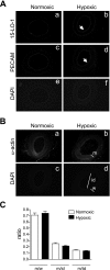
Immunohistochemical analysis of the mesenteric arteries from normoxic and hypoxic rabbits. A: localization of 15-LO-1 in the arteries. Arterial sections were costained for 15-LO-1 (a and b), PECAM (c and d), and nucleus (e and f). 15-LO-1 and PECAM expression are localized to the endothelium (white arrow). B: expression of smooth muscle cell (SMC) α-actin in the arteries (a and b). Nuclei were stained with 4,6-diamidino-2-phenylindole (DAPI; c and d). The external diameter of the hypoxic or normoxic arteries were 350–450 μm. Media thickness (M) was determined from the α-actin staining, and wall thickness (W), internal diameter (ID) and total diameter (TD; 2 × W + ID) were determined from the DAPI staining as shown in B,c and B,d. Several radial measurements (C) [M-to-W (M/W), M-to-ID (M/ID), and M-to-TD (M/TD) ratios] were calculated. Values represent means ± SE.
Fig. 6.
Fig. 6.
Effect of AA on membrane potential (Em) of SMCs of rabbit mesenteric arteries. Em was determined by impaling the SMCs of mesenteric arteries. Em was measured in presence of vehicle, Indo (10−5 M), Indo and phenylephrine (Phe; 10−7 M), or Indo, Phe, and AA (10−4 M). Effect of BW-755C (BW, 10−4 M ) alone or with Indo, Phe, and AA was also determined in hypoxic arteries. A: original Em tracings from SMCs of a normoxic or hypoxic artery. B: averaged Em values (n = 5 arteries) under various conditions. Values are means ± SE. ***P < 0.001 and **P < 0.01 compared with the values in the previous column; ##P < 0.01 compared with the corresponding value in the normoxic arteries.
Fig. 7.
Fig. 7.
Contraction of mesenteric arteries from normoxic or hypoxic rabbits. A: contractions to KCl (45 × 10−3 M). B: contractions to cumulative concentrations of Phe (10−7–10−4 M) in endothelium intact (+E) or denuded arteries (−E). C–E: endothelium-intact arterial rings were pretreated with Indo (10−5 M) and nitro-l-arginine (l-NA; 3 × 10⁻⁵ M), and contractions to cumulative concentrations of Phe (10−7–10−6 M; C), serotonin (5-HT; 10−7–10−4 M; D), or U-46619 (10−9–10−6 M; E) were determined. Contractions are expressed as tension in grams. Values represent means ± SE. ***P < 0.001, and *P < 0.05 compared with the corresponding values in normoxic arteries. **P < 0.01, normoxic +E compared with hypoxic +E. $$P < 0.01, normoxic −E compared with hypoxic −E.
Fig. 8.
Fig. 8.
Relaxations in mesenteric arterial rings from normoxic or hypoxic rabbits. Arterial rings were pretreated with Indo (10−5 M) and l-NA (3 × 10⁻⁵ M), precontracted with Phe (10−7–10−6 M), and relaxations to acetylcholine (ACh; A) or AA (B) were determined. Values represent means ± SE. ***P < 0.001 and **P < 0.01 compared the corresponding values in the normoxic arteries.
Fig. 9.
Fig. 9.
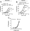
Effects of 15-LO-1 and small-conductance Ca2+-dependent K+ channel inhibitors on relaxations in mesenteric arterial rings from normoxic or hypoxic rabbits. Arterial rings were pretreated with Indo (10−5 M) and l-NA (3 × 10⁻⁵ M), and the inhibitors, precontracted with Phe (10−7–10−6 M) and relaxations to ACh, were determined. Effect of BW-755C (10−4 M) and nordihydroguaiaretic acid (NDGA; 2 × 10⁻⁵ M) on these ACh relaxations in normoxic (A) or hypoxic (B) arteries. Effect of apamin (2 × 10⁻⁷ M) on the ACh relaxations in normoxic (C) and hypoxic (D) arteries. Values represent means ± SE. ***P < 0.001.
Fig. 10.
Fig. 10.
Relaxations mediated by intermediate-conductance Ca2+-dependent K+ channels in mesenteric arterial rings from normoxic or hypoxic rabbits. Arterial rings were pretreated with Indo (10−5 M), l-NA (3 × 10⁻⁵ M), and vehicle, or various inhibitors, precontracted with Phe (10−7–10−6 M) and relaxations to ACh or 1-ethyl-2-benzimidazolinone (1-EBIO) were determined. ACh relaxations in the arteries from normoxic (A) and hypoxic (B) rabbits were incubated with Indo, l-NA, or 1-[(2-chlorophenyl)diphenylmethyl]-1H-pyrazole (TRAM-34; 10−5 M) in combination with vehicle or BW-755C (10−4 M). 1-EBIO relaxations in the arteries from normoxic and hypoxic rabbits (C) were incubated with Indo and l-NA. Values represent means ± SE. ***P < 0.0001 and *P < 0.05 for the effect of the inhibitors compared with the vehicle.

Comment in

  • Findings of research misconduct.
    [No authors listed] [No authors listed] NIH Guide Grants Contracts (Bethesda). 2013 Oct 25:NOT-OD-14-010. NIH Guide Grants Contracts (Bethesda). 2013. PMID: 24163871 Free PMC article. No abstract available.
  • Findings of Research Misconduct.
    [No authors listed] [No authors listed] Fed Regist. 2013 Oct 2;78(191):60873-60874. Fed Regist. 2013. PMID: 27737231 Free PMC article. No abstract available.

References

    1. Aalkjaer C, Lombard JH. Effect of hypoxia on force, intracellular pH and Ca2+ concentration in rat cerebral and mesenteric small arteries. J Physiol 482: 409–419, 1995. - PMC - PubMed
    1. Aggarwal NT, Chawengsub Y, Gauthier KM, Viita H, Yla-Herttuala S, Campbell WB. Endothelial 15-lipoxygenase-1 overexpression increases acetylcholine-induced hypotension and vasorelaxation in rabbits. Hypertension 51: 246–251, 2008. - PubMed
    1. Aggarwal NT, Gauthier KM, Campbell WB. 15-Lipoxygenase metabolites contribute to age-related reduction in acetylcholine-induced hypotension in rabbits. Am J Physiol Heart Circ Physiol 295: H89–H96, 2008. - PMC - PubMed
    1. Aggarwal NT, Holmes BB, Cui L, Viita H, Yla-Herttuala S, Campbell WB. Adenoviral expression of 15-lipoxygenase-1 in rabbit aortic endothelium: role in arachidonic acid-induced relaxation. Am J Physiol Heart Circ Physiol 292: H1033–H1041, 2007. - PubMed
    1. Bajpai AK, Blaskova E, Pakala SB, Zhao T, Glasgow WC, Penn JS, Johnson DA, Rao GN. 15(S)-HETE production in human retinal microvascular endothelial cells by hypoxia: novel role for MEK1 in 15(S)-HETE induced angiogenesis. Invest Ophthalmol Vis Sci 48: 4930–4938, 2007. - PubMed

Publication types

MeSH terms

LinkOut - more resources