Case Reports
. 2009 Jan;46(1):97-104.
doi: 10.1354/vp.46-1-97.

Clinico-pathologic features of fatal disease attributed to new variants of endotheliotropic herpesviruses in two Asian elephants (Elephas maximus)

Affiliations
Case Reports

Clinico-pathologic features of fatal disease attributed to new variants of endotheliotropic herpesviruses in two Asian elephants (Elephas maximus)

M M Garner et al. Vet Pathol. 2009 Jan.

Abstract

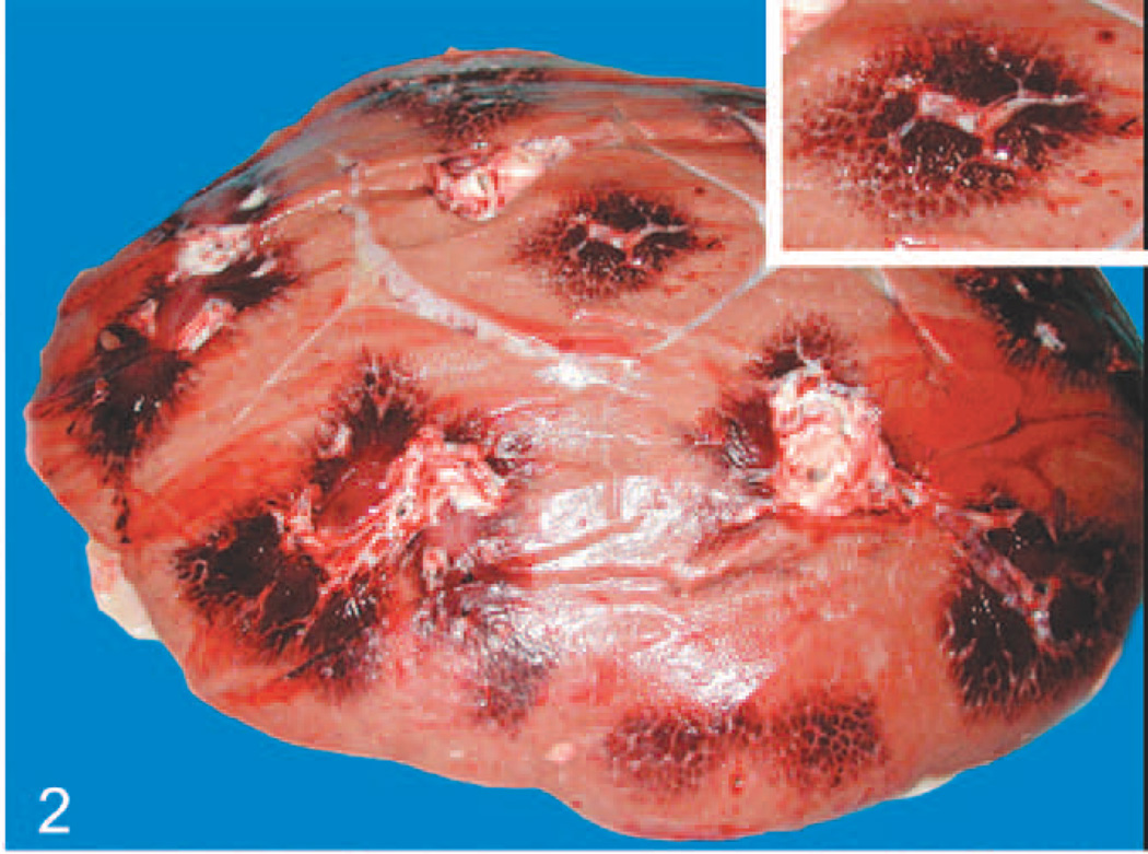
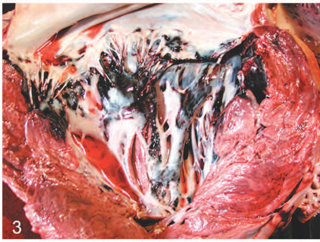
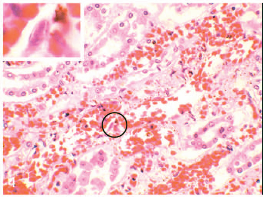
The first herpesviruses described in association with serious elephant disease were referred to as endotheliotropic herpesviruses (EEHV) because of their ability to infect capillary endothelial cells and cause potentially fatal disease. Two related viruses, EEHV1 and EEHV2, have been described based on genetic composition. This report describes the similarities and differences in clinicopathologic features of 2 cases of fatal endotheliotropic herpesvirus infections in Asian elephants caused by a previously unrecognized virus within the betaherpesvirus subfamily. EEHV3 is markedly divergent from the 2 previously studied fatal probosciviruses, based on polymerase chain reaction sequence analysis of 2 segments of the viral genome. In addition to ascites, widespread visceral edema, petechiae, and capillary damage previously reported, important findings with EEHV3 infection were the presence of grossly visible renal medullary hemorrhage, a tropism for larger veins and arteries in various tissues, relatively high density of renal herpetic inclusions, and involvement of the retinal vessels. These findings indicate a less selective organ tropism, and this may confer a higher degree of virulence for EEHV3.


Figures

Fig. 1
Fig. 1
Elephant No. 2. Note marked edema in mesentery root with associated petechiae and ecchymoses.
Fig. 2
Fig. 2
Elephant No. 2, kidney on cut surface shows lobular reddening, especially of the medullary regions, corresponding to congestion within and hemorrhage around the peritubular capillaries. Inset: Higher magnification of medullary reddening.
Fig. 3
Fig. 3
Heart; elephant No. 2. Note hemorrhage in left atrioventricular valve leaflets and associated endocardium, and to a lesser extent in the myocardium of the ventricular septum and left ventricular free wall.
Fig. 4
Fig. 4
Kidney; elephant No. 2. Note congestion of the medullary peritubular capillaries associated with some mild hemorrhage and scattered interstitial cellular debris. Inset: Higher magnification of circled region showing intranuclear amphophilic inclusion in an endothelial cell. HE.
Fig. 5
Fig. 5
Artery in splenic capsule; elephant No. 1. Note hypertrophy of endothelial cells, Some of which contain intranuclear inclusions (arrows). Inset: Higher magnification of circled endothelial cell showing intranuclear amphophilic inclusion. HE.
Fig. 6
Fig. 6
Liver; elephant No. 2. Note congestion and sinusoidal leukocytosis. Sinusoids also contain blue bodies (arrow), determined by electron microscopy to be degenerative inflammatory cells. Inset: Higher magnification of 2 blue bodies from circled region. HE.
Fig. 7
Fig. 7
Electron photomicrograph of artery from splenic capsule; elephant No 1. Note hypertrophy of endothelial cells and accumulation of electron dense granules in the cytoplasm of center cell, m = muscular tunic. Inset left: Note nonenveloped hexagonal particle in nucleus (n), budding of particle through nuclear membrane (arrow), and enveloped particles in the cytoplasm. Inset right: Intracytoplasmic enveloped hexagonal particles typical of herpesvirus, uranyl acetate and lead citrate. Bar =125 nm.
Fig. 8
Fig. 8
PCR detection of a novel herpesvirus POL gene in the heart tissue; elephant No. 1. Gel electrophoretic separation of first-round PCR products by using Codehops primers LGH2595/2597 (550 bp). Photograph of ethidium bromide stained agarose gel. Lanes 1 to 4, undiluted DNA samples from No. 1. Lane 1, whole blood; 2, heart; 3, intestine; 4, spleen; 5, EEHV1B positive control; 6, no DNA negative control.
Fig. 9
Fig. 9
PCR detection of EEHV3 TER gene in all tissues; elephant Nos. 1 and 2. Gel electrophoretic separation of first-round PCR products by using primers LGH6707/6708 (320 bp). Photograph of ethidium bromide stained agarose gel. Lanes 1, 2, 9, and 10, elephant No. 1, undiluted DNA samples; 1, intestine; 2, spleen; 9, whole blood; 10, heart. Lane 3, human placental DNA negative control. Lanes 4 to 8, elephant No. 2., undiluted DNA samples; 4, whole blood; 5, colon; 6, kidney; 7, liver; 8, spleen. Elephant No. 1 intestine was also positive (not shown).

References

    1. Burkhardt S, Hentschke J, Weiler H, Ehlers B, Ochs A, Walter J, Wittstatt U, Goltenboth R. Elephant herpes virus—a problem for breeding and housing of elephants. Berl Munch Tierarztl Wochenschr. 1999;112:174–179. (German) - PubMed
    1. Ehlers B, Burkhardt S, Goltz M, Ochs A, Weiler H, Hentschke J. Genetic and ultrastructural characterization of a European isolate of the fatal endotheliotropic elephant herpesvirus. J Gen Virol. 2001;82:475–482. - PubMed
    1. Ehlers B, Dural G, Marschall M, Schregel V, Goltz M, Hentschke J. Endotheliotropic elephant herpesvirus, the first betaherpesvirus with a thymidine kinase gene. J Gen Virol. 2006;87:2781–2789. - PubMed
    1. Fickel J, Liechfeldt D, Richman LK, Streich WJ, Hildebrandt TB, Pitra C. Comparison of glycoprotein B (gB) variants of the elephant endotheliotropic herpesvirus (EEHV) isolated from Asian elephants (Elephas maximus) Vet Microbiol. 2003;91:11–21. - PubMed
    1. Fickel J, Liechfeldt D, Reinsch F, Goritz F, Hildebrandt TB. Investigations on the occurrence of herpes virus infections in Asian elephants (Elaphas maximus) Adv Ethol. 2000;35:133.

Publication types

LinkOut - more resources