. 2009;60(3):741-9.
doi: 10.1093/jxb/ern319. Epub 2008 Dec 26.

Efficient mitochondrial targeting relies on co-operation of multiple protein signals in plants

Affiliations

Efficient mitochondrial targeting relies on co-operation of multiple protein signals in plants

Laurent Chatre et al. J Exp Bot. 2009.

Abstract

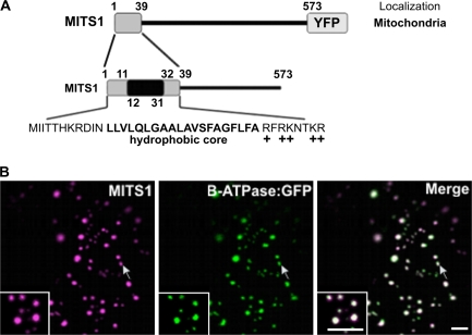
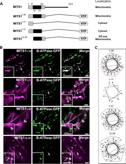
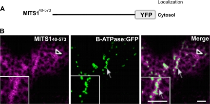
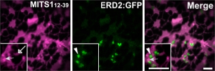
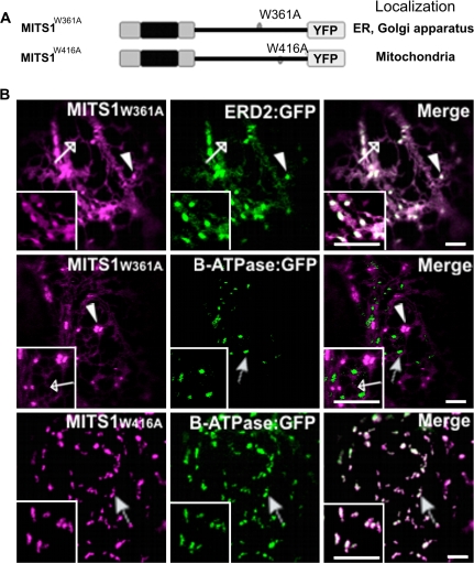
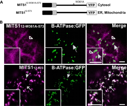
To date, the most prevalent model for transport of pre-proteins to plant mitochondria is based on the activity of an N-terminal extension serving as a targeting peptide. Whether the efficient delivery of proteins to mitochondria is based exclusively on the action of the N-terminal extension or also on that of other protein determinants has yet to be defined. A novel mechanism is reported here for the targeting of a plant protein, named MITS1, to mitochondria. It was found that MITS1 contains an N-terminal extension that is responsible for mitochondrial targeting. Functional dissection of this extension shows the existence of a cryptic signal for protein targeting to the secretory pathway. The first 11 amino acids of the N-terminal extension are necessary to overcome the activity of this signal sequence and target the protein to the mitochondria. These data suggest that co-operation of multiple determinants within the N-terminal extension of mitochondrial proteins may be necessary for efficient mitochondrial targeting. It was also established that the presence of a tryptophan residue toward the C-terminus of the protein is crucial for mitochondrial targeting, as mutation of this residue results in a redistribution of MITS1 to the endoplasmic reticulum and Golgi apparatus. These data suggest a novel targeting model whereby protein traffic to plant mitochondria is influenced by domains in the full-length protein as well as the N-terminal extension.


Figures

Fig. 1.
Fig. 1.
MITS1 harbours an N-terminal targeting signal and is localized to mitochondria. (A) Schematic representation of MITS1 and of its N-terminal region. Positively-charged residues follow a 20 residue hydrophobic region, characteristic of a mitochondrial targeting sequence. (B) In epidermal cells of tobacco leaves, MITS1:YFP labels punctate structures of various sizes that colocalize with the mitochondrial marker β-ATPase:GFP (arrows). Insets: magnified section of main panels. Scale bars=5 μm.
Fig. 2.
Fig. 2.
Exploration of the N-terminal 39 residues of MITS1 reveals that a co-ordination of three regions is required for efficient mitochondria targeting. (A) Schematic representation of the N-terminal consecutive domains fused to YFP and their subsequent intracellular localizations. (B) Region 1–39 efficiently targets a YFP to mitochondria (arrow) and the YFP punctate structures fully co-localize with β-ATPase:GFP (arrows). 1–11:YFP (missing the central hydrophobic core and the positively-charged region) and 1–31:YFP (missing the positively-charged region) were localized to the cytosol (empty arrowheads). (C) The Helical Wheel Projection of MITS1 N-terminal pre-sequence shows a cationic cluster in 1–39 and 12–39 sequences (but not in the other pre-sequence truncations) consistent with their localization to mitochondria (blue dots are hydrophobic residues, + indicates positive charge). Insets: magnified section of main panels. Scale bars=5 μm.
Fig. 3.
Fig. 3.
Residues 1–39 of the N-terminal extension are required for MITS1 to reach mitochondria. (A) A schematic representation of MITS1 lacking the first 39 amino acids. (B) In the absence of the N-terminal pre-sequence, MITS1 was found in the cytosol (empty arrowhead), which in plant cells assumes a diffuse yet reticulated appearance. No co-localization was noticed with the mitochondrial marker, β-ATPase:GFP (arrow). Insets: magnified section of main panels. Scale bars=5 μm.
Fig. 4.
Fig. 4.
MITS112–39 does not localize at Golgi bodies. To exclude the possibility that the punctate structures labelled by MITS112–39 peptide fusion were Golgi bodies, cells were cotransformed with the ER/Golgi marker, ERD2:GFP (Boevink et al., 1998) and MITS112–39. As shown in this figure, MITS112–39 labelled the ER (empty arrow), and dots (full arrow), which did not colocalize with the Golgi (arrowhead). These dots corresponded to mitochondria as shown in Fig. 2. Insets: magnified section of main panels. Scale bars=5 μm.
Fig. 5.
Fig. 5.
Tryptophan 361 influences the activity of the MITS1 N-terminal pre-sequence. (A) Schematics of the mutations within MITS1 fusions to YFP. (B) Confocal images of tobacco leaf epidermal cells coexpressing a MITS1:YFP mutant and either ERD2:GFP or β-ATPase:GFP. MITS1W361A:YFP was found in the ER (empty arrow) and Golgi apparatus (arrowhead) as confirmed by the ER/Golgi apparatus marker ERD2:GFP (ER, empty arrow; Golgi apparatus, arrowhead). The mutation of tryptophan 416 to alanine did not affect the distribution of MITS1 to mitochondria (arrow). Insets: magnified section of main panels. Scale bars=5 μm.
Fig. 6.
Fig. 6.
Tryptophan 361 mutation influences the behaviour of a truncated MITS1. (A) Schematic representation of the MITS112–573 constructs. (B) Confocal images of tobacco leaf epidermal cells show distribution of MITS112-W361A-573:YFP in the cytosol (empty arrowhead) but no colocalization with β-ATPase:GFP. MITS112–573 was found in the ER (empty arrows) and dots. Most of these colocalized with mitochondria (full arrows) but not with the Golgi (see Supplementary Fig. S1 at JXB online). Insets: magnified section of main panels. Scale bars=5 μm.

Comment in

References

    1. Addya S, Anandatheerthavarada HK, Biswas G, Bhagwat SV, Mullick J, Avadhani NG. Targeting of NH2-terminal-processed microsomal protein to mitochondria: a novel pathway for the biogenesis of hepatic mitochondrial P450MT2. Journal of Cell Biology. 1997;139:589–599. - PMC - PubMed
    1. Alder NN, Johnson AE. Cotranslational membrane protein biogenesis at the endoplasmic reticulum. Journal of Biological Chemistry. 2004;279:22787–22790. - PubMed
    1. Bannai H, Tamada Y, Maruyama O, Nakai K, Miyano S. Extensive feature detection of N-terminal protein sorting signals. Bioinformatics. 2002;18:298–305. - PubMed
    1. Batoko H, Zheng HQ, Hawes C, Moore I. A rab1 GTPase is required for transport between the endoplasmic reticulum and Golgi apparatus and for normal Golgi movement in plants. The Plant Cell. 2000;12:2201–2218. - PMC - PubMed
    1. Boevink P, Oparka K, Santa Cruz S, Martin B, Betteridge A, Hawes C. Stacks on tracks: the plant Golgi apparatus traffics on an actin/ER network. The Plant Journal. 1998;15:441–447. - PubMed

Publication types