. 2008 Dec;4(12):e1000317.
doi: 10.1371/journal.pgen.1000317. Epub 2008 Dec 26.

Ion channel clustering at the axon initial segment and node of Ranvier evolved sequentially in early chordates

Affiliations

Ion channel clustering at the axon initial segment and node of Ranvier evolved sequentially in early chordates

Alexis S Hill et al. PLoS Genet. 2008 Dec.

Abstract

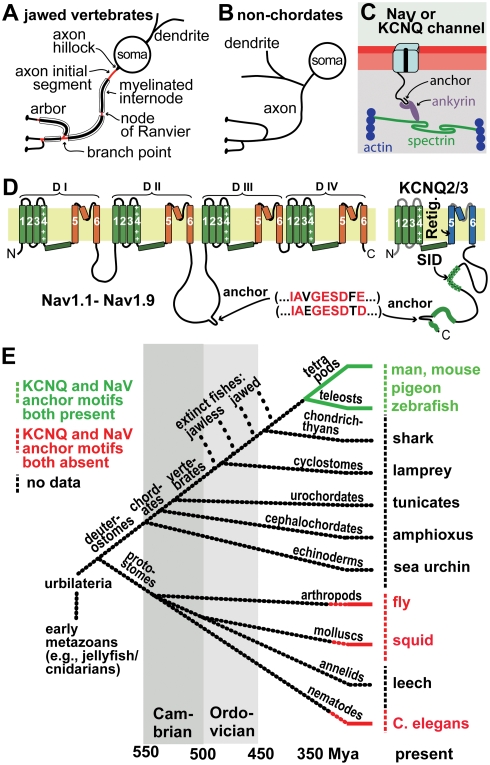
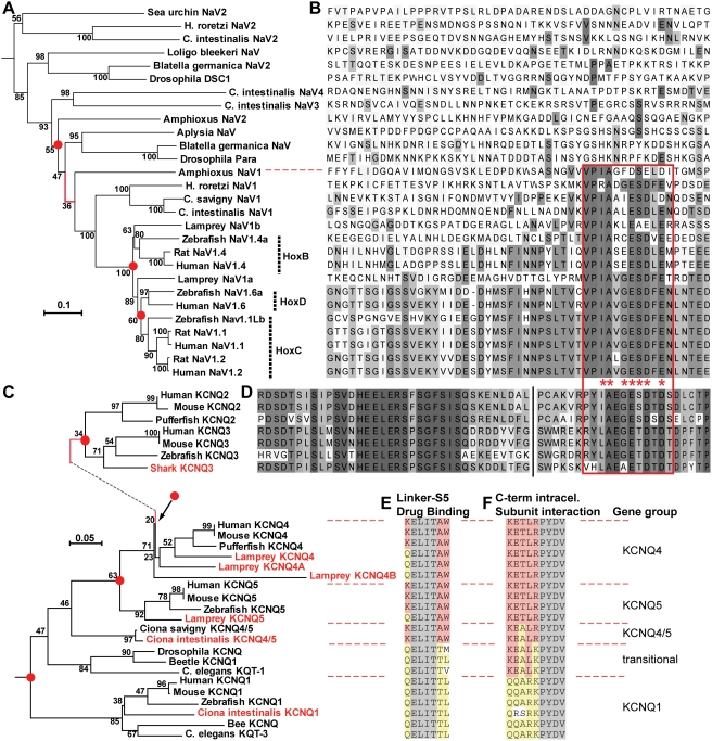
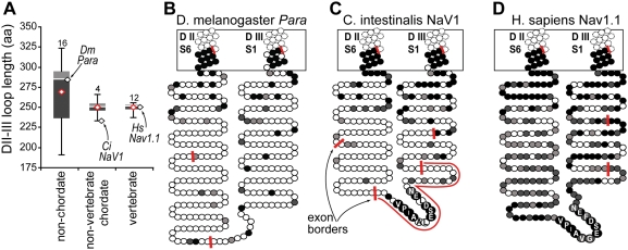
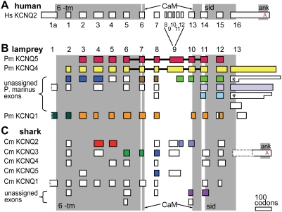
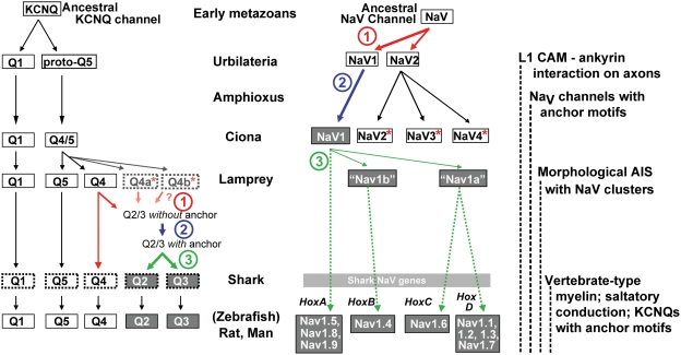
In many mammalian neurons, dense clusters of ion channels at the axonal initial segment and nodes of Ranvier underlie action potential generation and rapid conduction. Axonal clustering of mammalian voltage-gated sodium and KCNQ (Kv7) potassium channels is based on linkage to the actin-spectrin cytoskeleton, which is mediated by the adaptor protein ankyrin-G. We identified key steps in the evolution of this axonal channel clustering. The anchor motif for sodium channel clustering evolved early in the chordate lineage before the divergence of the wormlike cephalochordate, amphioxus. Axons of the lamprey, a very primitive vertebrate, exhibited some invertebrate features (lack of myelin, use of giant diameter to hasten conduction), but possessed narrow initial segments bearing sodium channel clusters like in more recently evolved vertebrates. The KCNQ potassium channel anchor motif evolved after the divergence of lampreys from other vertebrates, in a common ancestor of shark and humans. Thus, clustering of voltage-gated sodium channels was a pivotal early innovation of the chordates. Sodium channel clusters at the axon initial segment serving the generation of action potentials evolved long before the node of Ranvier. KCNQ channels acquired anchors allowing their integration into pre-existing sodium channel complexes at about the same time that ancient vertebrates acquired myelin, saltatory conduction, and hinged jaws. The early chordate refinements in action potential mechanisms we have elucidated appear essential to the complex neural signaling, active behavior, and evolutionary success of vertebrates.


Conflict of interest statement

The authors have declared that no competing interests exist.

Figures

Figure 1
Figure 1. Axonal ankyrin-dependent NaV and KCNQ2/3 channel clusters and anchor motifs: neuronal cellular and molecular features associated with jawed vertebrates and absent from non-chordates invertebrates.
(A, B) Cartoons showing characteristic jawed vertebrate (A) and non-chordate (B) types of neuronal polarity. Many jawed vertebrate neurons have myelinated axons, and axonal domains bearing ankyrin-dependent channel clusters, which mediate AP initiation and conduction (AISs, nodes, and branch points, red). Non-chordate dendrites and axons arise from a common neurite, and lack myelin and channel clusters. (C) Proposed molecular interactions between jawed vertebrate axonal NaV and KCNQ channels, ankyrin-G, spectrin, and actin. (D) Cartoons showing NaV and KCNQ2/3 channel topology. Locations of peptide sequences required for KCNQ opener interaction (Retig., retigabine), tetramerization (SID, subunit interaction domain), and the axonal anchor motif are indicated. (E) Cladogram showing some nomenclature and important evolutionary relationships among animals; timeline is approximate. At right are listed model species whose channel sequences were previously shown to lack anchor motifs (red) or bear them (green), and those newly studied here (black).
Figure 2
Figure 2. Phylogenetic analysis reveals that anchor motifs evolved sequentially in chordates (NaV channel) and jawed vertebrates (KCNQ2/3).
(A) Phylogram (minimal evolution) of NaV channels, showing that all vertebrate channels are derived from chordate NaV1. The branch on the phylogram in which the anchor motif first evolved is shown in red. Key nodes, associated with gene duplications, have red dots. Nodes are labeled with bootstrap values. (B) Alignment of NaV channel DII–DIII loop sequences, showing presence of anchor motifs in chordate NaV1 and all vertebrate channels (below dotted red line). The anchor motifs are boxed (red). Shading indicates each residue's conservation within the aligned 28 NaV sequences: bins represent ≤10, 11–20, 21–30, 31–45, 46–60, and 61–100% conservation. (C) Phylogeny of KCNQ channels, based on analysis of amino acids encoded on exons 5–7. Novel genes identified or cloned in this study are highlighted (named in red) As in A, key nodes associated with gene duplications are highlighted with red dots, and branch marking the inferred first appearance of the anchor motif is shown in red. (D) Alignment of KCNQ2 and KCNQ3 C-terminal intracellular sequences near the anchor motifs. Break (vertical black line) indicates location of 5–8 omitted, poorly conserved residues. The KCNQ2/3 anchor motif (red boxed region) is similar but non-identical to that of chordate NaV genes. Otherwise no homology to the NaV DII–DIII loop sequence shown in B is evident. Shading indicates conservation within the 7 KCNQ sequences aligned: shades represent ≤15, 15–30, 31–45, 46–60, 61–75, 76–90, and 91–100% conservation. (E–F) Aligned sequences at key functional sites for genes compared phylogenetically in C. Shading: grey, conserved in all KCNQ subunits; yellow, conserved in jawed vertebrate KCNQ1 subunits; red, conserved in jawed vertebrate KCNQ2-5 subunits. (E) Peptide sequence at the border of the S4-5 pore linker and the S5 pore helix, including (in KCNQ2-5 orthologues) the W residues required for retigabine interaction. (F) A portion of the tetramerization, or subunit interaction, domain. Scale bars: substitutions per residue.
Figure 3
Figure 3. The NaV channel DII–III intracellular loop is poorly conserved in invertebrates lacking the anchor motif, and highly conserved in vertebrates.
(A) Plot showing lengths of DII–III loop sequences of NaV channels, deduced from cDNA clones. Stick bars show range, grey boxes show 2nd and 3rd quartiles, and red diamond shows average length. Black diamonds show lengths of loops from species indicated. (B–D) Cartoons depicting the degree of sequence conservation and exon borders (red bars) of orthologous NaV channels from D. melanogaster (para), C. intestinalis (Nav1), and H. sapiens (Nav1.1) in the region between D II S6 and DIII S1. Each shaded circle is one amino acid. In non-chordates (e.g., fly), the transmembrane and very membrane-proximal portions of the intracellular loop show high conservation with vertebrates, but the remainder of the loops are poorly conserved in sequence and length. In protochordates (e.g., C. intestinalis), a series of highly conserved residues (VPIAAIESDLDN, residues labeled) appears on a short, novel exon (red line in C); the rest of the loop is poorly conserved like other invertebrate genes. However, the mean length of the 4 known protochordate NaV1 loops is nearly identical to those of vertebrates. Among vertebrate genes (e.g., human Nav1.1), the entire loop is more highly conserved, and has a simplified exon structure, with the anchor motif part of the same, exceptionally long exon as the conserved DII6 transmembrane segment. The shading scheme is based on alignment of the indicated sequence and six vertebrate NaV channel sequences. Shading scale represents, from darkest to lightest, matching of 5–6 of 6, 3–4 of 6, 2 of 6, and 0–1 of 6 vertebrate sequences.
Figure 4
Figure 4. NaV immunostaining of lamprey brain and spinal cord reveals linear profiles similar in appearance to mammalian AISs.
(A) Transverse cryosection through lamprey spinal cord immunolabeled for NaV channels (yellow). Nuclei are stained using DAPI (blue). Large distal Müller and Mauthner axons show little NaV channel membrane immunolabeling, but small intensely labeled profiles have morphology suggestive of AISs, and are clustered near the motor column. Red lines and box indicate approximate location, plane and orientation of adjoining higher magnification horizontal (Bi, Bii) and transverse (Biii) section images. (Bi) Dorsal sensory neuron, with a bipolar axon. Both rostral and caudal axon branches show increased NaV channel immunolabeling in their proximal portions (arrows). (Bii) AIS-like profiles are abundant in oblique horizontal sections near grey matter. (Biii) Higher magnification view of AIS-like NaV channel immunostaining near motor column in spinal cord cross-section. (C) Low power view of lamprey rhombencephalon in whole mount. Reticulospinal neurons have been back-filled via their large descending axons. Somata, narrowed initial segments, and large distal axons of Müller and Mauthner cells are indicated. Box encloses the location shown at higher magnification in panel D. (D) Widefield epifluorescence image of lamprey rhombencephalon whole mount showing soma and AIS of Mauthner neuron immunolabeled for NaV channels (yellow). Scale bars: A, 125 µm; Ai, 20 µm; B, Bi, Bii, 25 µm, Biii, 12.5 µm.
Figure 5
Figure 5. Lamprey motor system axons have narrow initial segments with NaV channels clusters.
(A) Detail of lamprey left rhombencephalon region whole mount showing large reticulospinal Mauthner (Mth) and Müller (Mu) neurons, backfilled via their spinal axons by in vivo FITC-dextran injection (green), then fixed and immunostained against NaV channels (mouse Pan NaV, red). AISs of two Mth and Mu neurons are marked (arrowheads). (B) Higher magnification view of red-boxed region in A, showing NaV channel immunolabeling at membrane of Mauthner neuron AIS. (C) Lamprey spinal cord whole mount showing several motoneurons filled in vivo via their distal axons with FITC-dextran (green), then fixed and immunostained against NaV channels (red). (D) Higher magnification view of red-boxed region in C, showing dense clustering of NaV channels at narrow proximal AIS of a motoneuron axon. Scale bars: 40 µm (A), 10 µm (B), 20 µm (C), 10 µm (D).
Figure 6
Figure 6. C. intestinalis KCNQ1 is more prominently expressed in neurons than is KCNQ4/5.
Subunit mRNA expression was detected using whole mount in situ hybridization. Animals were allowed to develop at 18°C for the indicated times after fertilization in vitro, then labeled with antisense RNA probes for C. intestinalis KCNQ1 (A–C) or C. intestinalis KCNQ4/5 (D–F), and stained using NBT/BCIP. (A) At 10.5 hours post-fertilization, a pair of tail dorsal midline neurons are stained (arrowhead). (B) At 11.6 hours post-fertilization, numerous dorsal and ventral epidermal sensory neurons in tail and trunk (arrowheads, left), and labeling of the cerebral ganglion (right), is apparent. (C) At 17.2 hours post-fertilization, continued staining of central and peripheral neurons of free swimming larva is apparent C1. Strong staining of caudal portion of cerebral ganglion (arrowhead). C2. Staining of epidermal sensory neurons (arrowheads). (D) At 10.5 hours post-fertilization, KCNQ4/5 staining is strongly apparent in the notochord, but absent from central and peripheral neurons. (E) At 11.6 hours post-fertilization, strong notochord staining persists, and weaker staining of ventral cerebral ganglion is detectable. (F) At 15.5 hours post-fertilization (immediately before hatching), weak staining is detected in the posterior-ventral half of the cerebral ganglion. eTB, early tailbud; mTB, mid-tailbud; lTB, late-tailbud; hL, hatched larva. Scale bar, 100 µm.
Figure 7
Figure 7. C. intestinalis KCNQ4/5 gives small currents in Xenopus oocytes, but forms heteromers with mammalian KCNQ3 that express more efficiently.
(A) Family of large KCNQ1 currents elicited by voltage steps. (B) Family of small C. intestinalis KCNQ4/5 currents elicited by voltage steps. (C, D, F, G, H) Co-expression of C. intestinalis KCNQ4/5 with rat KCNQ3 results in expression of heteromeric currents with altered kinetic properties. Expression of rat KCNQ3 only resulted in currents (not shown) undistinguishable from uninjected oocytes (E). Co-expression of C. intestinalis KCNQ4/5 with rat KCNQ3 produced currents that were larger in amplitude than C. intestinalis KCNQ4/5 alone (C, F), activated at more depolarized membrane potentials (D, G), and had steeper voltage-dependence (H).
Figure 8
Figure 8. The KCNQ ankyrin-interaction domain evolved in the transition between ancestral jawless and jawed vertebrates.
(A) Human (H. sapiens, Hs) KCNQ2 exon structure, numbered based on previous reports . Grey boxes indicate locations of functionally conserved domains (6TM, the six transmembrane segments and pore region; CaM, the discontinuous calmodulin-binding IQ domain; sid, the subunit interaction domain mediating tetramerization; ank, the conserved domain containing the ankyrin-interaction motif). (B) Diagram summarizing lamprey (P. marinus, Pm) KCNQ genomic analysis and cDNA cloning indicating that lampreys possess KCNQ1, KCNQ5, KCNQ4, and, possibly, two additional KCNQ4-like genes. Exons (renumbered as indicated) linked in silico by overlapping of genomic sequencing traces are shown in identical colors. Exons linked by cDNA cloning are connected by heavy black bars. Unlinked exons are shown in white. Two different exon 1 traces had start codons that could not be determined (due to poor conservation, dotted borders). KCNQ1 exons were confirmed by reciprocal BLAST analysis versus vertebrate and invertebrate genomes. Five different non-KCNQ1 3′ exons (exon 13) were identified; two were represented in the genomic traces by sequences with different stop codon positions (asterisks). This may be the result of heterozygosity in the source genomic DNA . (C) Diagram of shark (C. milii, Cm) KCNQ gene family as elucidated from the partially sequenced genome. Exons containing orthologues of mammalian KCNQ1 through KCNQ5, identified by reciprocal BLAST search, are indicated. One trace contained the ankyrin binding domain (distal exon 13 region) of KCNQ3.
Figure 9
Figure 9. Anchor motifs evolved sequentially in NaV and KCNQ channel families.
Diagrams summarize the evolutionary history of KCNQ channels (left), NaV channels (right), and their anchor motifs. In each gene family, three steps are highlighted: (step 1, red arrows) gene duplication preceding appearance of the anchor, (step 2, blue arrows) evolution producing the anchor motif, and (step 3, green arrows) additional duplication resulting in parologues conserving the motif. Representative species studied are listed in the center. Genes possessing anchor motifs are shaded grey. The NaV channel motif arose before the common ancestor of amphioxus and tunicates. In KCNQ channels, an inferred KCNQ2/3 gene acquired the motif, after lamprey but before the duplication producing shark KCNQ2 and KCNQ3. Where 3 or more genes are shown arising from an ancestor gene, an unresolved sequence of gene duplications (i.e., polytomy) is present. Genes apparently lacking orthologues in more recently evolved phyla are indicated by asterisks. Genes identified genomically without cDNA confirmation have dashed border boxes. Lamprey KCNQ4a/b genes are drawn lightly, indicating their uncertain status (see Results). Shark NaV genes (not characterized in this study) are omitted. Hox-linked vertebrate NaV genes underwent lineage-specific genome duplications, as indicated by boxed gene groups. Associated hox clusters are labeled ,. Ankyrin interaction with L1 CAMs on axons evolved before the deuterostome-protostome divergence –.

Comment in

References

    1. Hille B. Ionic channels of excitable membranes. Sunderland, Mass.: Sinauer; 2001.
    1. Schweigreiter R, Roots BI, Bandtlow CE, Gould RM. Understanding myelination through studying its evolution. Int Rev Neurobiol. 2006;73:219–273. - PubMed
    1. Hartline DK, Colman DR. Rapid conduction and the evolution of giant axons and myelinated fibers. Curr Biol. 2007;17:R29–35. - PubMed
    1. Rolls MM, Satoh D, Clyne PJ, Henner AL, Uemura T, et al. Polarity and intracellular compartmentalization of Drosophila neurons. Neural Develop. 2007;2:7. - PMC - PubMed
    1. Ramon y Cajal S. Texture of the nervous system of man and the vertebrates. New York: SpringerWein; 1907. Pasic P, Pasic T, translators and editors. 2002.

Publication types

MeSH terms